Classification of infectious diseases. Classification of Infectious Diseases
L. 15 Es. inf. diseases.doc
Lecture 15.MAIN GROUPS OF INFECTIOUS DISEASES.
1. Clinic and prevention of dysentery in school
Dysentery - A type of intestinal infection. The disease is manifested by a lesion of the mucous membrane of the large intestine with a dysentery stick, while there are painful sensations and intoxication phenomena.
The causative agents of dysentery are bacteria belonging to the entero-typhoid group: sticks Sonne, FlexnerGrigorieva - Shigi, UnionSchmitzet al. These bacteria are immobile due to the lack of movement organs; they are highly resistant in the environment: in the discharge of the patient they persist up to 48 hours, and in winter - up to 100 hours. Dysenteric sticks die in the sunlight for 30 minutes, when heated to 50-60 ° C - for 10 minutes, in solution phenol (1%) - within 30 minutes.
The source of dysentery is a sick person, the discharge of which contains pathogens. The sick person is dangerous from the very first days of the illness, since from his hands the pathogenic microbes get on everyday objects and as a result can get on food and water. That is why dysentery is sometimes called “dirty hands disease”.
Pathways of dysentery are similar to the pathways of other intestinal infections - contact and domestic transmission of infection against the background of personal hygiene violations. Microbes enter the human body through the mouth (mainly with water). An important role in the spread of infection is played by flies, which carry dysenteric sticks on their paws from cesspools, toilets, from landfills to food.
Outbreaks of dysentery are seasonal: occur in the autumn and summer months.
Pathogens enter the colon and linger in the folds of the mucous membrane. Here they feed, excrete toxins, and multiply. As a result, the intestinal mucosa is damaged: swelling, redness, bleeding, mucus secretion, ulceration. The waste products of pathogenic microbes - toxins - spread through the blood throughout the body, affecting organs and systems.
The first symptoms of dysentery occur in 3-5 days. Depending on the severity of the disease, dysentery can be easymoderate and severe (toxic).
With moderate formsymptoms manifest suddenly. Body temperature rises to 38-38,5 ° C. The patient's condition deteriorates rapidly: weakness, lethargy, general malaise develops, appetite decreases or disappears altogether; nausea, vomiting are possible. The stool becomes liquid. After 1-2 days, the stool acquires a typical form of dysentery: mucopurulent discharge with streaks of blood. The chair quickens, accompanied by stabbing cramping pain, often in the left half of the abdomen. On examination, the abdomen is somewhat swollen, painful on pressure, on the left, a tense sigmoid colon is felt. The amount of mucus increases rapidly, the stool loses its fecal character and its volume decreases significantly. These symptoms persist for about a week, after which the general condition of the patient improves, the body temperature decreases. The number of bowel movements per day is reduced and the stool becomes more decorated, but for another 1-2 weeks it can be unstable, keeping the mucous and bloody look.
Treatment.Treatment of patients with dysentery involves their mandatory isolation. The patient is recommended rest, compliance with mandatory hygiene measures. Antimicrobials are used for seven days and, if necessary, longer. During the treatment period, the patient is prescribed a special diet, abundant drinking, with a toxic form, intravenous administration of drugs and nutrients is used. Current disinfection is required at the bedside.
^ Prevention of dysentery. Preventing dysentery in school is one of the necessary measures for the localization of the disease.
When a child with dysentery is identified, an immediate isolation and examination of persons who are in direct contact with him is needed.
The school premises are disinfected: the classroom, the kitchen, the toilet, the lobby, the gym, and others. The catering staff is examined for bacilli carrier.
Children who have had dysentery attend classes only after permission from the doctor.
The measures to prevent the occurrence and spread of dysentery in school include:
maintaining cleanliness of classrooms;
compliance with the rules of storage and trade in food products;
maintenance of public toilets;
the destruction of flies;
timely emptying of garbage cans;
strict control over the personal hygiene of schoolchildren;
removal of children from cleaning bathrooms, washing places
general use, garbage collection and food waste;
proper organization of drinking regime at school;
familiarizing students and their parents with symptoms
and the danger of dysentery.
^ 2. Clinic and prevention of viral hepatitis in school
Hepatitis - an acute infectious disease affecting mainly the liver tissue, which causes the pathology of the liver functions and against this background - a metabolic disorder in the body.
Hepatitis can be primaryand in this case it is an independent disease, or secondary, then it is a manifestation of another disease. Development secondary Hepatitis is associated with exposure to hepatotropic factors - viruses, alcohol, drugs or chemicals.
Viral hepatitis (Botkin's disease) is a viral disease characterized by general poisoning of the body with the products of vital activity of pathogenic viruses. At the same time, there is a violation of skin pigmentation (icteric color), an increase in the size of some internal organs (spleen, liver).
Exists two types of pathogens viral hepatitis - viruses types A and B. Hepatitis A bears the name infectiousfoothepatitis A, hepatitis B - whey. The source of viral hepatitis is a sick person or a virus carrier. The maximum infectiousness of the patient falls on the preicteric period of the disease and the first days of the icteric period.
For different types of hepatitis vary and transmission mechanisms infections. Causative agents hepatitis A from the blood of a virus carrier or patient fall into its discharge, and then household contact - In the body of a healthy person. Causative agents hepatitis b are found only in the blood of the patient, where they persist for a long time after his recovery. Infection with hepatitis can occur. with the patient's blood: through various medical instruments, blood transfusions or intrauterine.
The incubation period for infectious hepatitis is 7-45 days; serum hepatitis - 2-6 months. During this period, there are no external manifestations of the disease.
Course of the disease two periods:
preicteric (more than 1 week). The main symptoms of this period are general weakness, malaise, loss of appetite, feeling of heaviness in the epigastric region, belching, short-term fever, pain in the joints, in the liver. At the end of this period, there is a change in the color of the discharge: the urine becomes brown and the feces turn white;
icteric (2-4 weeks). The main manifestations of this period are a change in the color of the soft palate and later - of the skin (yellowing); the appearance of itching of the skin. Against this background, the size of the liver and spleen increase, sometimes there is bleeding of the skin and mucous membranes.
Hepatitis B is more severe than hepatitis A. Hepatitis B is characterized by constantly growing weakness, aversion to food, the appearance of multiple vomiting, sleep disturbance, a sharp icteric staining of the skin and visible mucous membranes, and pruritus.
^ Hepatic coma- complication of hepatitis B - one of the main causes of the lethal outcome of the disease. In addition, the disease can become chronic, during which periods of exacerbations occur.
^ Treatment of viral hepatitis. The patient is prescribed bed rest - in this position the blood supply to the liver improves. Due to the decrease in blood clotting due to hepatitis B, the risk of bleeding of various kinds increases, so caregivers need to be careful when injecting.
Patients with hepatitis A are given a special diet with a restriction on the amount of fat in their diet, the exclusion of protein from it and an increase in the content of amino acids. A large amount of fluid is also prescribed.
With symptoms of intoxication, intravenous drip infusion of 5% glucose solution, hemodez up to 1 - 1.5 l. At the same time, hormone therapy is prescribed (prednisone, hydrocortisone). Also apply ointment, relieves itching.
In the event of the appearance of symptoms of hepatic coma, hormonal preparations and antibiotics are used.
It is important to remember that the patient is a source of infection, so it is necessary to disinfect his laundry, dishes, and discharge. Sanitary cleaning of the room should be carried out at least three times a day.
^ Prevention of viral gepa titus in school includes:
Information about the absence of students in the classroom is transmitted to medical points.
The educational institution is notified of all cases of illness of students and their families.
Students who have missed school for more than three days are allowed to attend classes only with the permission of the doctor.
There is an explanatory work with students and their parents about the danger, symptoms and measures of prevention of viral hepatitis.
Strict control is exercised over school employees, especially catering workers.
Compliance with sanitary and hygienic requirements, rules of transportation and storage of food products, etc. is strictly checked.
Active work is being carried out to identify the sick,
who have no symptoms of the disease.
All cases of the disease are reported to the sanitary
epidemic services.
All school premises are disinfected (especially toilets).
If necessary, quarantine is declared.
^ 3. The most common childhood intestinal infections.
Typhoid and paratyphoid A and B. These are acute infectious diseases of a bacterial nature related to intestinal infections. Their pathogens withalmonella - mobile short gram-negative sticks with rounded ends. All three pathogens are fairly stable in the external environment - in water, soil, food, household items, they persist for 2-3 months.
The source of the disease are sick or carrier. The greatest number of pathogens is excreted in the midst of the disease along with feces and urine. The patient becomes infectious from the first days of the disease and remains so even in the last days of the incubation period, and in some cases after recovery. The contagiousness of a person can last from several months to decades.
Ways tocottages of typhoid paratyphoid diseases fetidal-oral character Transmission is through water, food, contaminated hands, household items, etc.
In typhoid fever, the latent incubation period of the disease ranges from 7 to 20 days, an average of 14 days, with paratyphoid fever from 3 to 14 days, an average of 7-8 days.
^ Symptoms and course. Typically, the course of typhoid paratyphoid diseases is cyclical: the first 5-6 days there is a gradual increase in painful symptoms, the temperature rises to 39-40 ° C; headache, insomnia, weakness, blackout, delirium appear on the 4-5th day; tongue lined, skin rash, pulse rare, uneven; On the 7th-8th day, the main symptoms and complications appear (intestinal bleeding, perforation of ulcers of the intestines with subsequent peritonitis, otitis, mumps, psychosis, meningitis). In the future, there is a weakening of the clinical manifestations of the disease and a gradual decrease in temperature: the last period of the disease begins - convalescence (normalization of the condition, lowering of the temperature).
The diagnosis of typhoid paratyphoid diseases is made on the basis of a blood test from a vein and a bacteriocarrier examination by planting stools and urine.
Treatment.For the treatment of patients with typhoid paratyphoid diseases, the antibiotics chloramphenicol and syntomycin are used. It is important that the patient observe bed rest and a strict diet. Due to heavy bleeding, the patient needs constant drinking and regular meals, even in the absence of his appetite.
^ Prevention of all intestinal infections (see dysentery and infectious hepatitis A).
Another way to prevent an epidemic of typhoid and paratyphoid fever is by timely vaccinationswhich are used for epidemic indications. Vaccinations are carried out with a high incidence in the area or with the threat of an outbreak of the disease. The following vaccines are used for immunization:
Killed vaccine - it is administered subcutaneously in 0.5 and 1 ml twice with an interval of! 0-14 days;
Chemical vaccine - administered once a dose of 1 ml.
Repeated vaccinations (revaccination) are carried out in a year
Once by both vaccines in a dose of 1 ml.
Food toxicoinfection.
These are diseases caused by pathogens belonging to groups salmonella, staphylococcus (streptococcus) and opportunistic microorganisms. Infection occurs as a result of taking infood products.
Salmonella caused by ingestion salmonell - very resistant microorganisms that persist in food for a long time. Main ispoint of infection - animals (cattle, dogs, pigs) and birds (especially waterfowl). A person falls ill with salmonellosis by eating meat, milk or eggs of sick animals and birds.
The first signs of salmonella disease appear after 12–24 hours. Its symptoms are nausea, vomiting, headache, abdominal pain, diarrhea, convulsions, and a drop in heart activity. The body temperature is normal or slightly elevated, in severe cases, high. The severity of the disease depends on the type of pathogen and the number of germs ingested. Duration of illness - 3-5 days. Severe forms of this disease can be fatal.
The diagnosis is made on the basis of data of epidemiological analysis and examination, acute onset and rapid development of symptoms of the disease, as well as laboratory tests.
Treatment.Mild forms of salmonellosis do not require treatment. For severe forms, medical assistance is needed, which consists in washing the stomach and then restoring the activity of the systems affected by the disease. Heat, hot water bottles to the feet or a warm bath are recommended as auxiliary means for raising the heart activity.
With staphylococcal food poisoningpoisonous substances are formed in food - enterotoksiny,characterized by thermal stability and antigenic properties.
Botulism - food poisoning caused toxin botulinum sticks. The causative agent of botulism is a mobile sporiferous bacillus (bacillus), which lives in anaerobic conditions and produces a poison - exotoxin, which is in the first place in toxicity. Its disputes are widespread in the external environment and are highly resistant.
Source of infections are warm-blooded animals, in the intestines of which botulinum sticks inhabit. Human infection occurs through various products, in which the pathogen multiplies and toxin accumulates. Especially dangerous in this regard are cannednye products from fish and vegetables.Diseases of botulism are also common as a result of mushroomimproperly mothballed at home.
The first symptoms of the disease occur about a day after ingestion of products infected with botulinum bacillus. At the same time, there are signs of damage to the central nervous system, manifested by nausea, general weakness, dizziness, headache and double vision, dilated pupils, difficulty speaking and swallowing.
Treatment:immediate gastric lavage and the introduction of a special serum. Serum therapy is recommended to be combined with the use of biomitsin.
^
4. Acarosis and helminthiasis in children.
Acarose
Scabies - a contagious disease caused by scabexact itch (mite). Infection cause only females, because after fertilization, the male dies, and the female penetrates the skin, where it runs parallel to its surface mangemove. Lays there eggs. After 5-6 days, the immunity hatches out through the roof of the course. After 3-7 weeks, young mites become sexually mature.
Outside the body of the "owner" at a temperature of 60 ° C mites die within an hour. Ticks and larvae almost immediately die when boiling and ironing with a hot iron. Ticks die when treated with 3% carbolic acid, creolin, xylene; eggs are more resistant to these and other disinfectants.
Source of infection with scabies is sick person. There are direct and indirect infection routes scabies. With direct paths (from 41 to 95% of patients), the pathogen passes directly from a sick person to a healthy person at the time of bodily contact. Indirect the path is the transmission of the pathogen through household items, primarily personal use.
The incubation period for scabies is from 8 days to 16 weeks.
SymptomsItchy skin - the main subjective symptom of the disease, while it can be defined as strong, moderate, weak or painful. Itching increases in the evening and at night and is the first symptom of the disease. The main diagnostic sign of scabies, distinguishing it from other diseases, is considered scabby - a slightly elevated straight or curved line of whitish or off-white color 5-10 mm long. At the front (head) end of the entrance there is a female tick in the form of a dark dot. Localizationscabby passages - on the hands, wrists, palms, on the elbows, on the mammary glands of women, on the genitals of men, less often in the navel, abdomen, thighs, chest, etc.
Treatment.Sent to the destruction of the pathogen with the help of some chemicals: sulfur ointment, ointment Wilkenson, soap "K", etc.
Prevention.When identifying patients with scabies in an organized children's team, it is necessary to conduct anti-epidemic measures in the outbreak: children should be barred from attending school and kindergartens for the duration of the full treatment. All patients who have been in contact with patients should be identified, examined and medical observation should be established. An entire class may be subject to preventiveskys treatment, if several cases of scabies are registered in it or if “fresh” cases of scabies are detected during the observation of the lesion. A major role is played by the organization of the current and final disinfectionwhich aims to destroy the pathogen in the environment. Disinfection of bedding, towels, underwear is made by boiling in 1-2% soda solution or any laundry detergent for 5-10 minutes from the moment of boiling. Outerwear (dresses, suits, trousers, sweaters, etc.) are ironed on both sides with a hot iron, paying attention to the pockets. Some things (coats, raincoats, fur coats, leather goods, etc.) can be rendered harmless by hanging in the open air for 5 days. When disinfecting some things (soft toys, shoes, outerwear), you can temporarily exclude them from use on 5-7 days by placing in a separate plastic bag. A wet cleaning with a 1-2% soap-soda solution is performed daily in the patient’s room.
Lice are transmitted mainly by direct contact orthrough bedding, wearing clothes. They can be carriers of infectious diseases (typhus, relapsing fever, etc.). The spread of lice is observed with insufficient hygienic care, uncleanliness, crowding, i.e. with a low material and cultural level of the population. There are pediculosis of the head, pediculosis of the trunk, pediculosis of the pubis.
^ Treatment and prevention. When lice of the head hair is washed with soap, combed out with a thick comb the remaining lice and nits. After that, you need to wash your hair with warm vinegar, as it dissolves the chitinous cover of nits. Apply and other tools available in a pharmacy that are recommended by a doctor.
If body lice are detected, it is necessary to apply frequent soap baths, rub the affected areas with 5% boric ointment or 2% boron tar paste. In the presence of ploshchits skin is treated with a warm solution of table vinegar or shave pubic hair and rubbed with 5-10% white or gray mercury ointment.
Prevention of lice is achieved by increasing the material and cultural level of the population, the dissemination of sanitary and hygienic knowledge, and observance of individual prophylaxis (regular body washing, bed linen change, hair care, and a control inspection of linen for possible lice under unsatisfactory sanitary conditions, etc.) .
Helminthiasis
Ascariasis . Ascaris - large dioecious helminth from the class of roundworms. The length of the female is 25-40 cm, and the male is 15-25 cm, the body is spindly, reddish. The female lays a day more than 200 thousand eggs, released from the human intestine with feces. These eggs are still non-infectious, they must undergo development in the external environment under favorable conditions: temperature 24-26 ° С, moist soil, access of oxygen. In the absence of moisture and in direct sunlight, they quickly die. The development of ascaris from the larva to the first laying of eggs lasts 9-15 weeks.
Source of infection is only sick a person is 2.5-3.5 months after infection and remains so for more than six months. Directly from an infected person, infection of the people around him does not occur, since eggsmust be developed in the external environment. The mechanism of transmission of ascariasis - fecal-oral. Transmission factors are soil, vegetables, dirty hands.
SymptomsThe larvae pierce the intestinal mucosa, penetrate the bloodstream and spread to all organs. The first phase of the disease is a dry cough, asthmatic, or symptoms of bronchitis, or flu, or "urticaria." In the second phase of the disease, a disorder of the CNS occurs. Ascaris exchange products irritate the nerve endings of the intestine. This explains the violation of the functions of the stomach, spasmodic bowel obstruction. General intoxication, disorder of appetite, sleep, moods, disobedience, decrease in progress.
Prevention.Strict implementation of hygiene skills: thorough washing of hands after going to the toilet and working in the garden, washing fruits and vegetables (wean children from the bad habit of nail biting). It is very dangerous to use fresh feces as a fertilizer in gardens and kitchen gardens, they must be composted.
Enterobiasis. Pathogen - pinworm. This is a small (5-12 mm) helminth from the class of roundworms. Live in lower section fineintestine, cecum and in the initial section of the large intestineperson Females descend into the rectum at night, youcrawling out of the anus, laying eggs in its folds and dying. The life span of the helminth is 4 weeks. Eggs ripen in 4-6 hours, mature pinworms actively move, causing itching in the anus. When combing the eggs fall into the subungual space, contaminated bedding and underwear and household items. This causes both self-infection and infection of other people.
A source infection - a person secreting helminth eggs. Infection occurs by ingestion of eggs in food, water and on dirty hands.
SymptomsThe first symptoms of the disease is itch in the regiontee anal fold in the evening and night. The secondary infection gets to the place of scratching, causing inflammatory processes and weeping eczema. Then there may be cramping abdominal pain, rapid stool with mucus, tenesmus. Functional disorder of the central nervous system is manifested by the same symptoms as in ascariasis.
Prevention.Implementation of sanitary and hygienic measures (see ascariasis). An infected child should be washed with warm water and soap daily in the morning and evening. Daily iron hot sheets and panties. In case of ascariasis and enterobiasis, it is necessary to consult a doctor and undergo a course of drug treatment.
Whipworm infestation occurs as a result of the ingestion of eggs when using contaminated vegetables, “fruits, berries, water, as well as when putting eggs into the mouth with dirty hands. Susceptibility to whipworm universal. The worm is quite widespread in many regions.
A school is not only a “temple of knowledge,” but also a place where large numbers of people gather. In the face of infection, the school can become one of the foci of the disease. This is due to the fact that children, not yet possessing a strong and stable immunity, do not always adequately fulfill the requirements of personal hygiene. Teachers and parents need to remember: how well children are accustomed to look after themselves depends on their security from the occurrence of infectious diseases.
Each student must comply with the following personal hygiene rules.
Daily make morning toilet.
Wash hands thoroughly before eating and after each use of the toilet.
Do not put foreign objects in your mouth: pen, pencil, etc .; when reading books do not slobber fingers.
Keep your workplace clean and tidy.
Eat only in specially designated
this places (if available), etc.
regularly air the classrooms;
daily wet cleaning of classrooms and corridors of the school;
check for replacement shoes for students, especially
in the autumn and spring periods;
antibacterial treatment of toilets;
to observe the hygienic cleanliness of food processing units;
to control food.
^ 6. Especially dangerous infections.
Especially dangerous infections are characterized by high virulence and pathogenicity.
Plague - Acute infectious disease belonging to the group of zoonoses. Source of infections are gnawus (rats, gophers, gerbils, etc.) and sick person. Disease proceeds in forms bubonic, septic(rarely) and pulmonary. The most dangerous pulmonary form of the plague. Causative agent of infection - the plaguenaya wand, stable in the environment, well tolerating low temperatures.
There are two types of natural foci plague: foci of "wild", or steppe, plague and rat foci, urban or portthe plague.
Ways of Transmission plague is associated with the presence of insects (fleas, etc.) - transmissible. In case of pneumonic plague, the infection is transmitted. cartstuffy drip by (by inhalation of droplets of sputum of a sick person containing the causative agent of the plague).
Symptoms diseases manifest suddenly three days after infection, with a strong intoxication of the whole organism. Against the background of severe chills, the temperature quickly rises to 38-39 "C, there is a severe headache, facial hyperemia, the tongue is covered with white bloom. In more severe cases, delusions of hallucinatory order, cyanosis and sharpness of facial features develop with the appearance of suffering on it, sometimes horror Quite often, in every form of plague, diverse skin phenomena are observed: hemorrhagic rash, pustular rash, etc.
With bubonicthe form of plague that usually occurs when a flea is bitten, the cardinal symptom is the bubo, which is an inflammation of the lymph nodes.
Secondary development septicforms of the plague in a patient with the bubonic form can also be accompanied by numerous complications of a non-specific nature.
Primary pulmonarythe form is the most dangerous in the epidemic and very severe clinical form of the disease. Its sudden onset: body temperature quickly rises, coughing and excessive sputum appear, which then becomes bloody. In the midst of the disease characteristic symptoms are general depression, and then an excited-delusional state, high fever, signs of pneumonia, vomiting with blood, cyanosis, shortness of breath. Pulse quickens and becomes filiform. The general condition is deteriorating dramatically, the patient's strength is dying out. The disease lasts 3-5 days and ends in death without treatment.
Treatment.Treatment of all forms of plague is made with the use of antibiotics. Streptomycin, terramycin and other antibiotics are prescribed individually or in combination with sulfonamides.
Prevention.In natural foci, observations are made on the number of rodents and carriers, their examination, disinfestation in the most threatened areas, examination and vaccination of healthy populations.
A special role in the fight against plague is given to the timely detection of the first cases of the disease, the immediate isolation and hospitalization of patients. All persons in contact with the patient, contaminated things and the corpse of a person who died from the plague, are also isolated for six days. Conducted emergency prophylaxis with antibiotics of all in contact with the patient. Quarantine is imposed on the settlement in which the patient is identified. It is forbidden to leave the population.
Vaccinationis carried out dry live vaccine subcutaneously or percutaneously. The development of immunity begins from the 5-7th day after a single injection of the vaccine.
Cholera - acute intestinal infection, characterized by the severity of the clinical course, high mortality and the ability in a short time to bring a large number of victims. Causative agent of cholera - vibrio cholerae, which has a curved shape in the form of a comma and has great mobility. Recent cases of cholera outbreak are associated with a new type of pathogen - vibrio El tor.
The most dangerous way of spreading cholera is waterway. This is due to the fact that Vibrio cholerae can be stored in water for several months. Cholera is also characteristic of fecal-oral mechanism transfer.
The incubation period for cholera is from several hours to five days. It may be asymptomatic. There are cases when, as a result of the most severe forms of cholera disease, people die in the first days and even hours of illness. The diagnosis is made using laboratory methods.
Main the symptoms cholera: sudden watery, profuse diarrhea with floating flakes, reminiscent of rice water, turning into a mushy and later in loose stools, profuse vomiting, decreased urination due to fluid loss, leading to a state in which blood pressure drops, pulse becomes weak, there is a strong shortness of breath, cyanosis of the skin, tonic convulsions of the muscles of the extremities. The features of the patient's face are sharpened, the eyes and cheeks are sunken, the tongue and mucous membrane of the mouth are dry, the voice is hoarse, the body temperature is lowered, the skin is cold to the touch.
Treatment:massive intravenous administration of special saline solutions to compensate for the loss of salts and fluids in patients. Assign antibiotics (tetracycline).
^ Control and prevention measures. To eliminate foci the disease is carried out a complex of anti-epidemic measures: through the so-called "courtyard bypasses", patients are identified, the persons who have been in contact with them are isolated; Provisional hospitalization of all patients with intestinal infections, disinfection of foci, control over the good quality of water, food and their neutralization, etc. are carried out. If there is a real danger of the spread of cholera, as a last resort quarantine.
With the threat of disease, as well as in areas where there are cases of cholera, immunization population killed by cholera vaccine subcutaneously. Immunity to cholera is short and not high enough; therefore, after six months revaccination is carried out by a single injection of a vaccine in a dose of 1 ml.
anthrax - typical zoonotic infection. The causative agent of the disease is thick immobile wand (bacillusla) - has a capsule and spore. Anthrax spores persist in the soil for up to 50 years.
A source infections - homemadethe animals, cattle, sheep, horses. Sick animals excrete the pathogen with urine and feces.
^ Ways anthrax spreads are diverse: contact, food, transmissive (through the bites of blood-sucking insects - gadfly and flies).
The incubation period of the disease is short (2-3 days). By clinical forms distinguish dermal, gastrointestinaland pulmonary anthrax.
With dermalanthrax forms a stain first, then a papule, a vesicle, a pustule, and an ulcer. The disease is severe and, in some cases, fatal.
With gastrointestinalform of the predominant symptoms are sudden onset, a rapid rise in body temperature to 39–40 ° C, acute, cutting abdominal pain, bloody vomiting with bile, bloody diarrhea. Usually, the disease lasts 3–4 days and most often ends in death.
Pulmonarythe form has an even more severe course. It is characterized by high body temperature, impaired activity of the cardiovascular system, a strong cough with the release of bloody sputum. After 2-3 days, the patients die.
Treatment.The most successful is the early use of specific anthrax serum in combination with antibiotics. When caring for the sick, it is necessary to observe personal precautions - work in rubber gloves.
Preventiondiseases include the identification of sick animals with the appointment quarantine, disinfection of fur clothing for suspected infections, immunization according to epidemic indicators.
Smallpox. It is an infectious disease with cartstuffy drip the infectious mechanism of transmission. Causative agent of smallpox - virus "Taurus Pashen - Morozov", having a relatively high resistance in the external environment. Source of infection - sick person during the entire period of the disease. The patient is infectious within 30-40 days, until the complete collapse of the smallpox crusts. Infection is possible through clothing and household items with which the patient came into contact.
The clinical course of smallpox begins with an incubation period of 12-15 days.
Three possible forms smallpox: easy form - varioloid or smallpox without rash; smallpox ordinary type and drain smallpox, severe hemorrhagic a form that occurs in cases of hemorrhages in the elements of the rash, as a result of which the latter become purple-blue ("black pox").
^ Easythe form of smallpox is characterized by the absence of rash. General lesions are mild.
Smallpox ordinary typesuddenly begins with a sharp chill, rising body temperature to 39-40 ° C, headache and sharp pains in the sacrum and lower back. Sometimes this is accompanied by the appearance on the skin of a rash in the form of red or red-purple spots, nodules. The rash is localized in the inner thighs and lower abdomen, as well as in the pectoral muscles and the upper inner part of the shoulder. The rash disappears in 2-3 days. In the same period, the temperature decreases, the patient feels better. After that, a smallpox rash appears, which covers the whole body and the nasopharyngeal mucosa. At the first moment, the rash has the character of pale pink dense spots, on top of which a bubble forms ( pustule). The contents of the bubble gradually becomes cloudy and suppurate. During the period of suppuration, the patient feels a rise in temperature and acute pain.
Hemorrhagicthe form of smallpox (purpura) is severe and often ends in death 3-4 days after the onset of the disease.
Treatmentbased on the use of specific gamma globulin. Treatment of all forms of smallpox begins with the immediate isolation of the patient in a box or private room.
Preventionsmallpox consists in the complete vaccination of children from the second year of life and subsequent revaccination. As a result, cases of smallpox almost never occur.
If diseases of smallpox occur, they conduct revaccination of the population. Persons who were in contact with the patient are isolated for 14 days in a hospital or in a temporary hospital deployed for this purpose.
^
7. The most common children's airborne infections.
Flu. This is an airborne infection, the most intense infectious disease. Periodically, influenza occurs in the form of large epidemics, during which in large cities from 30 to 70% of the population is ill.
Causative agent of influenza - viruscharacterized by variability. There are four types flu virus: A, B, C and ABOUT. In recent years, identified virus A variantswhich got the name A1 and A2. Those who have had the flu will develop immunity only to the virus that caused the disease. The resulting immunity is short-lived. The flu virus dies at room temperature.
A source infections - sick person. The incubation period for influenza ranges from several hours to 2-3 days.
Symptoms diseases: chills, fever, loss of appetite, headache, malaise, muscle pain. The disease lasts for 2-3 days, with the exception of severe toxic flu.
The recognition of influenza is difficult due to the fact that many infectious diseases have similar symptoms. For the diagnosis of influenza resort to laboratory tests.
Treatment.In the treatment of influenza, a number of agents are used to prevent complications, including antibiotics - penicillin, chloramphenicol, from sulfanilamide drugs - norsulfazole. A good therapeutic effect in recent years has been obtained with the introduction of specific gamma globulin.
Preventionthe flu is immunized with live influenza vaccine, as well as serum. In children's institutions for the preventive purpose, specially made gamma globulin is used.
Acute respiratory viral infections (ARVI). Caused by various viruses (adenoviruses, enteroviruses, etc.). Sources, modes of transmission, symptoms, etc., see flu).
Chickenpox - a disease from the group herpes virusinfectionswhich also includes herpetic disease, cytomegaly, mononucleosis.
A source diseases sick a person who becomes contagious in the last hours of incubation (6-8 hours). The entire period of the rash and 5 more days after the rash is dangerous. The transmission mechanism for chickenpox is airborne. People’s susceptibility to chicken pox is very high. When an outbreak in a kindergarten occurs, almost everyone who has not been ill before, becomes ill. As a result of the disease is produced persistent lifelong immunity. Localization of the infection process - in the epithelial cells of the mucous membranes of the respiratory tract, eyes, nose and skin.
The incubation period is 11-21 days.
SymptomsThe main symptom of the disease is a rash. Rash appears on the body from the 2nd day of illness, gradually covers all parts of the body, even the scalp, oral mucosa, pharynx, etc. A rash appears in the form of stains (roseol)that go into papules (tubercles)then in vesicles and puspurulent contents. On the 5-7th day, the rash dries out and peels off, complications are rare.
Prevention.Chickenpox is the most common infectious disease (after flu and acute respiratory infections) among children. It refers to “unmanaged” infections, because the transmission mechanism is extremely active, and specific prevention is not used. The main attention is paid to preventing the introduction of the virus into the children's team and the formation of the outbreak. Patients are isolated to the stage of falling off of all crusts. Children who did not have chickenpox, if they were in contact with patients, should be isolated for 21 days.
Scarlet fever. Causative agent scarlet fever is hemolytic toxigenic streptococcus.
Source of infection is sicksince the onset of the disease.
Scarlet fever occurs most often in children infected with toxigenic streptococcus and not having antitoxic immunity.
The mechanism of transmission of the disease is aerogenic, the transmission paths are airborne and airborne dust. Scarlet fever can be transmitted through toys and patient care items.
Immunity is produced in those who have been ill (antitoxic).
The incubation period ranges from 3 to 7 days.
SymptomsThe main localization of the infection process is pharynx, less commonly skin. Start acute, with high temperature rise. The patient complains of sharp pains in the throat, vomiting attacks are possible. The characteristic manifestations of scarlet fever include rash and sore throat.
A rash appears a few hours after the temperature rises. It is shallow, on a red background, consistently covers the trunk, cheeks, neck, ears (armpits, inguinal folds, flexor surfaces of the elbows and knees). The duration of the preservation of rash and peeling in the same sequence of 3-7 days.
The leading symptom is anginathat appears on
2-4thillness day and may manifest in necrotic form.
At the same time, the language has a very distinctive appearance: it is first coated, and then becomes crimson.
Complications of scarlet fever - rheumatism, rarely otitis, nephritis, lymphadenitis.
Prevention.Anti-epidemic measures - isolation of the patient for 22 days, quarantine for 7 days. Enhanced disinfection mode. Disinfection of toys, dishes, items for patient care. A vaccine against scarlet fever has not been developed. There is a specific gamma globulin, which is administered by contact within 5 days after isolation of the patient. Examination of children - daily.
Diphtheria. Causative agent of diphtheria - diphtheria stick, resistant to drying and low temperatures, which ensures its safety at various objects (toys, food products), but is quite sensitive to high temperatures (dies at 58-60 ° C) and the effects of disinfectants. Diphtheria sticks by their ability to produce exotoxin divided into 2 groups: toxic and non-toxic.
Source of infection is sick clinically or asymptomatic or (more often) bacteria carrier. The term of infectiousness is calculated from the last days of the incubation period until complete recovery (about 20 days). The main localization of the pathogen is on the mucous membrane of the upper respiratory tract, which determines the airborne and contact-everyday mechanism of transmission. In addition to diphtheria pharynx distinguish diphtheria croupdiphtheria nose and places of rare localization - eyes, vulva, skin (these forms are transmitted exclusively by contact and household).
The incubation period is 2-7 days.
SymptomsThere are 2 forms diseases: non-toxic and toxic. With toxic form Against the background of general malaise, pain occurs when swallowing. The pharynx is hyperemic, a yellowish-gray bloom forms on the tonsils, which can also cover the small tongue, the arms, and the posterior pharyngeal wall. With a severe toxic form of the disease, the films penetrate into the larynx and into the nose, the body temperature reaches 39-40 ° C. From the 2-3rd day of the disease, swelling of the pharynx and subcutaneous cervical tissue is increasing. As a consequence of the process, laryngeal diphtheria can develop - true diphtheria croup. On the background of high fever appears rough barking coughand then noisy breathing, hoarseness and aphonia, up to asphyxia, when the blue and blue lips come on (cyanosis), convulsions begin. The child at this stage recovers rarely. Most frequent complications - myocarditis, paralysis of the limbs, deafness.
Prevention.A patient with diphtheria is hospitalized for the full course of treatment. The group in which a case of diphtheria is registered is subject to quarantine for 7 days. Persons in contact with the patient must undergo a single bacteriological examination. For children and staff medical monitoring is established for 7 days with daily inspection and thermometry.
The main effective preventive measure is vaccination. Currently, the following vaccine preparations are produced: adsorbed pertussis-diphtheria-tetanus toxoid (DTP), adsorbed diphtheria-tetanus toxoid (ADS) and anti-diphtheria serum for treatment. According to the vaccination schedule, primary immunization with DPT vaccine in children from 3 months of age is carried out three times with an interval of 1.5 months. Revaccination is carried out once in 1.5-2 years with anatoxin ADS. Modern vaccination calendar provides for revaccination of DPT (“Vaccination schedule”).
Whooping cough. Whooping cough - a disease caused by bacteria coclyusha. Sticks outside the human body for more than two hours do not live.
Source of the disease is sick. The greatest danger to others is in the catarrhal period of the disease (from the beginning - 2 weeks). In the stage of convulsive cough, the patient is still contagious, but not more than 2 weeks.
Transmission of the pathogen is carried out by airborne droplets, and infection is possible only with direct communication. With pertussis, passive immunity of newborns is practically not observed, therefore the person is susceptible to the stick from the first days of life (children at preschool age are especially susceptible).
After the disease is formed lifeimmunityRepeated cases of the disease are rare.
The incubation period is from 7 to 14 (21) days.
SymptomsLocalization of the infectious process - in the respiratory tract: larynx, trachea, bronchi. Act toxin causes spasm of the bronchi and respiratory muscles, in some cases - the defeat of the respiratory center.
^ Catarrhal period. The initial signs of the disease are characterized by a slight fever, a runny nose, and a slight cough. This stage lasts about 2 weeks.
^ Period of convulsive cough. The disease goes into stage
attacks of spasmodic cough, when a few cough shocks are followed by a wheezing breath and then an irresistible cough (up to 20 times). At the same time, the face becomes red, swollen,
vomiting and even involuntary urination can join. Attacks during the day can be repeated 5-50 times. This
the stage sometimes lasts up to 6 weeks.
^ Period of permission. It lasts for 1-3 weeks, until
all symptoms of the disease will disappear.
Measles. Measles is an infectious disease caused by a virus. It is stable in the environment, does not tolerate drying, but is very contagious.
Only a source infections - sick person. It is dangerous to others from the last two days of incubation, during the catarrhal period and the first days after the rash. The term infectiousness - 8-10 days. Susceptibility to measles in non-diseased and unvaccinated people is absolute. The measles transmission mechanism is airborne. Have recovered produced persistent life time immunizationtet. Localization of the infectious process - in the cells of the mucous membranes of the upper respiratory tract.
The incubation period is 8-10 days, it can last up to 17 and 21 days (for vaccinated).
SymptomsCatarrhal period begins with a rise in temperature (38-39 ° C), which decreases after 3-4 days. During this period, cough, runny nose, conjunctivitis, and photophobia are observed against the background of general malaise. On 2-3rdday of catarrhal period small white nodules with a red border appear on the mucous membrane of the cheeks (Belskiy-Filatov spots). On the 4th day of illness with repeated rise in temperature appears spotted rash in a certain sequence within 3 days: 1st day - behind the ears, on the face and neck, 2nd day - rash covers the body, 3rd day - limbs, from the 4th day the rash disappears in the same sequence.
Measles are more severe in children under 2 years of age. In vaccinated children, the disease is atypical, but it is an epidemic hazard. Complications: false croup (laryngism), pneumonia (50%), purulent otitis (25%), blepharitis.
Prevention.Patients are treated mainly at home, isolating from others for a period of illness up to 5 days, counting from the onset of the rash. Quarantine in children's institutions is set to 17 or 21 days when gamma globulin is administered to contact persons. Disinfection measures are not required due to the low resistance of the pathogen. For vaccination, live measles vaccine is used, which gives a reliable and long-lasting immunity.
Rubella - disease caused by rubella virus. The virus is unstable in the external environment.
A source diseases - sick, dangerous from the 1st day during the entire period of the disease, as well as on the 5-7th day after recovery. The general term of infectiousness is 10-12 days. The transmission mechanism is carried by airborne droplets. But possible intrauterine infection. Rubella is dangerous during pregnancy, especially in the first 16 weeks: the virus crosses the placenta and infects the embryo. Children more often ill 2-10 years. Localization of the virus - skin and lymphatic system person
The incubation period for rubella is from 11 to 22 days.
SymptomsThere is a slight short-term rise in temperature without catarrhal phenomena. Observed limfadenite - increase most often posterior cervical and occipital lymph nodes up to the size of a pea. They become tight and painful. 2-3 days after that appears rash consistently on the face, neck, throughout the body (mainly on the extensor muscles, back, buttocks). It lasts for 2-3 days and disappears without pigmentation and peeling.
Prevention.Isolation of patients from children's institutions is carried out, but does not give effect because of the infectiousness of patients during the incubation period and the presence of asymptomatic forms. Children in contact with patients are not subject to isolation.
Parotitis. Mumps is an acute infectious disease caused by a virus, unstable in the environment, highly contagious.
A source infections - sick starting from the 1st day of illness. The term infectiousness - 5-7 days. The virus is excreted with saliva, mucus from the nasopharynx. The transmission mechanism is predominantly airborne, but it can also be transmitted through infected objects (toys). Have been ill formed lastingimmunity. Localization process: parotidgland, pancreas and gonads.
The incubation period is 14-20 days with fluctuations from 11 to 23 days.
SymptomsThe onset of the disease is acute, general malaise, pain on the sides of the neck, swelling of the salivary glands, initially noticeable in front of the ear, then extending behind the ear and down. Soreness increases with chewing, swallowing, talking. The temperature is not typical, it can be subfibril or reach 38-40 ° C. The febrile period lasts 3-4 days, and in severe cases it takes 6-7 days.
Prevention.Of great importance in the fight against parotitis are the timely detection and isolation of patients for 10 days. Quarantine for an organized team is usually 21 days. The main anti-epidemic event is the vaccination of live mumps vaccine (VHF).
Polio. The causative agent of polio - ente virusroviruses. It tolerates freezing and drying, dies quickly when exposed to high temperatures and disinfectants. For a long time (up to 3-4 months) it can be stored in milk, water, wastewater, on contaminated linen and feces.
Source of infections are virusosteli and sick a person from the 1st day of illness, he is contagious for 1.5 months or more. The most infectious is the first week of the disease. The transmission mechanism is mainly fecal-oral, but may also be airborne, as the virus begins to be released from the body with nasopharyngeal mucus 30–40 hours after infection. After suffering the disease remains long immunity, but only to one type of pathogen. Localization of the pathological process: the virus penetrates from the nasopharynx or intestines into the blood, and then into the central nervous system and affects the motor nuclei of the spinal cordwhat causes paralysis of the lower and upper extremities.
The incubation period is 3-21 days.
SymptomsThere are the following forms diseases:
asymptomatic withlack of clinical manifestations, diagnosed only by laboratory method;
non-paralytic,from the 2-3rd day, the temperature rises, meninge symptoms appear, and in some cases the pressure of the cerebrospinal fluid rises. After 2-4 weeks, ends with recovery;
paralytic,acute, temperature 38.5–40 ° C, catarrhal symptoms of the nasopharynx, gastrointestinal disorders, lethargy, drowsiness or insomnia, signs of meningitis, hyperesthesia. From 2-5th day of illness paresis and paralysis muscles of the lower extremities (58-82%) are most often affected, less often is a combination of paralysis of the muscles of the trunk, neck and other parts of the body. Paralyzes are sluggish and asymmetrical, there is a limitation in movements, a decrease in muscle tone, a decrease or extinction of tendon reflexes. 1-2 weeks after the onset of paralysis develops amyotrophy. Recovery begins a few days after the onset of paralysis. After 4-6 months, the recovery process slows down and can last up to 2-3 years. Residual phenomena are characterized by the presence of atrophy of individual muscle groups, and therefore appear curvature, deformation and contracture of the limbs and trunk.
Prevention.Timely detection and isolation of suspected patients with poliomyelitis in the infectious diseases hospital. Discharge from the hospital no earlier than 40 days from the onset of the disease with clinical recovery. In the outbreak carried out the current and final disinfection. Vaccination.
^
8. Sexually transmitted diseases (STDs).
Infectious diseases transmitted from person to person, predominantly sexually - STDs or STDs (sexually transmitted diseases), are common, especially in recent years. Currently, there are more than 20 sexually transmitted pathogens, including bacteria, viruses, protozoa, yeast fungi, and arthropods. The diseases caused by them are characterized by high contagiousness, relatively rapid spread among certain groups of the population. Previously, these diseases were called venereal diseases, and according to the new World Health Organization (WHO) classification - sexually transmitted diseases or diseases.
In Russia, there has been an increase in the incidence of STIs since 1990. In the previous decade (1980–1990), the incidence rate was 5% per 100 thousand population. In 1994, the number of cases of syphilis only increased from 8 thousand (in 1980) to 127 thousand. The most alarming moment in this statistic is the increase in the incidence rate among children. If the incidence of syphilis among the adult population during this period increased by 16 times, among children it is almost 21 times. Of the 761 cases of childhood syphilis, registered in 1995, 502 children aged 11–14 years contracted sexually on their own, and not from their parents — household ones, as in previous years. The leaders of the Ministry of Health and Social Development of the Russian Federation see one of the reasons for such an incredible increase in the incidence among children in the absence of an adequate level of sex education among children and teenagers.
According to WHO, every tenth person in the world, including children and old people, is sick with this or that STD. In 2006, 2.0 million STD patients were registered in Russia.
^ Classification of sexually transmitted diseases (WHO)
According to the classification proposed by WHO, sexually transmitted diseases include 22 diseases, which are divided into 3 groups.
Syphilis.
Gonorrhea.
Shankroid
Lymphogranulomatosis inguinal.
Granuloma venereal.
Genitourinary chlamydia.
Genitourinary trichomoniasis.
Candida vulvovaginitis and balanoposthitis.
Genitourinary mycoplasmosis.
Genital herpes.
Genital warts.
Genital contagious mollusk.
Gardnerella vaginitis.
Urogenital shigellosis homosexuals.
Pubic pediculosis.
Scabies.
Infection caused by human immunodeficiency virus (AIDS).
Hepatitis B.
Cytomegaly.
Amebiasis (mainly in homosexuals).
Giardiasis.
Sepsis of newborns.
Classic venereal diseases
Syphilis. Chronic venereal disease characterized by damage to the skin, mucous membranes, internal organs, bones and nervous system.
^ Ways of transmission.The causative agent of syphilis - pale treponema(spirochaete)enters the human body through damaged epithelial mucous membranes during sexual contact with a sick person. Occasionally, infection can occur through close household contact; children become infected through the milk of a sick mother. Intrauterine infection of the fetus was noted, as well as infection at the time of delivery when the fetus passes through the birth canal of the mother infected with syphilis. So, in 2006, 25 cases of congenital syphilis were recorded in Moscow.
SymptomsSyphilis is characterized by a wavelike pattern with a change in the periods of rise in incidence and its decline.
^ Hidden period - This is the period from the moment of infection to the first symptoms of the disease. On average, it lasts a month, but may be longer (if the patient takes antibiotics in the treatment of other infections or in less weakened people).
^ Primary period syphilis begins with the appearance on the site of the introduction of the pathogen erosion or ulcers (hard chancre). The second symptom is an increase in nearby lymph nodes ( inguinal lymphadenitis). The duration of the first period is 1-1.5 months.
^ Secondary period begins with the appearance on the skin and mucous membranes of roseolous, papular and pustular eruptions ( rash), pigmentation is disturbed, hair falls hard, hoarseness is noted, all lymph nodes are enlarged. This period can last from 2-5 up to 10 years. Internal organs can be affected: liver, kidneys, nervous and endocrine systems, sensory organs, joints, bones.
^ Tertiary period characterized by education gum and hillocks and occurs 10-15 years after infection. He is characterized by more severe damage to internal organs.
Along with the described stages of syphilis sometimes observedlong asymptomatic course. Immunity after the transfer of syphilis is not produced. Diagnosis of syphilis is carried out:
by biological seeding, the discharge from chancre ulcers and
from syphilitic elements;
serological blood tests for antibodies
against the pathogen (Wasserman reaction).
Gonorrhea. This is an infectious disease of the mucous membranes of the urinary organs, as well as the mucous membrane of the oral cavity and rectum after orogenital or homosexual contacts. Pathogen: gonococcus fixed diplococcus, having the form of coffee beans, stable in the body against phagocytosis. Infection occurs almost exclusively through sexual contact. In some cases, it is possible for non-sexually infected girls to wash their genitals with sponges, infected secretions from sick parents, as well as when sharing a bed with bedding, with a sick bed, and when washing together in the bathroom. Infection of newborns can occur during childbirth, during the passage of the sick mother through the birth canal.
In recent years, the incidence has increased among young people (persons aged 15-19 years), who often have repeated infections. Attention is drawn to the fact of the constant increase in the incidence of women aged 16-18 years. At the present stage, there is a high proportion of patients with asymptomatic forms of gonorrhea. The percentage of simultaneous damage to the urinary organs by gonococci and other microorganisms, in particular chlamydia, viruses, mycoplasmas, trichomonads, fungi, etc., is increasing.
Symptoms Anatomical and physiological features of male, female and child organisms determine the specificity of the course of gonorrhea, therefore emit male, female and childgonorrhea. The latent period is usually 3-5 days.
Have a husbandrank first of all, the urethra is affected, which is expressed in cutting pains when urinating, profuse purulent discharge, in frequent irresistible urge to urinate.
Have of women gonorrhea occurs without subjective symptoms, but with damage to almost all parts of the urogenital system, as well as the rectum. The urethra, the vagina, the uterus, the appendages, the cervix, the ovaries, the circulatory tissue are affected.
Have girls mainly affects the vagina, urethra and anus. The uterus and appendages are not affected. Complaints are usually absent, occasionally there is a slight pain during urination and defecation.
Complications of gonorrhea occur in the form of gonococcal stomatitis, lesions of the oral mucosa, larynx, pharynx, eyes, kidneys, joint inflammation, infertility.
Diagnosis of gonorrhea is carried out by bacteriological examination of a smear of purulent discharge and bacteriological seeding with the aim of isolating a pure gonococcal culture.
The treatment of patients should be comprehensive with regard to comorbidities, individual, simultaneous for sexual partners.
Other sexually transmitted infections
A. With predominant lesion of the genitals
This group of infections includes sexually transmitted diseases, the causative agents of which are bacteria, virusses, protozoa, mushrooms and arthropods. Often these infections can accompany syphilis and gonorrhea. Practitioners point out that all of these infections occur. sluggish, they do not have an acute period, but immediately become chronic and stand out already when treating patients with infertility, miscarriages, inflammatory diseases of the urogenital system and internal organs.
Trichomoniasis - the most common disease of the urogenital tract . He ranks 3rd after gonorrhea and chlamydia. More often, young people are sick at the age of 20-30 in the period of maximum manifestation of the generative function and the greatest sexual activity. In women, the vagina is affected, and in men, the urethra is affected. There is itching, frothy discharge grayish-yellow color with an unpleasant odor. Untreated trichomoniasis gives up to 60% of complications - more often than with gonorrhea.
Genitourinary Mycoplasmosis cause mycoplasma. The disease proceeds covertly, affecting the vagina, urethra, cervix, with periodic sensations of itching in the urinary organs, mucous secretions and provokes the occurrence of pathology of pregnant women, fetuses and newborns (pneumonia).
Gardnerellosis causes rod bacteriumaffecting the urinary organs of men and women, as well as infectious processes in newborns (meningitis, pneumonia). Gardnerellosis is often diagnosed in women who have sex partners. There is a mixed infection with chlamydia, gonococci, trichomonads, fungi and anaerobic pathogens.
Genitourinary herpes . The most common infection during sexual intercourse or kissing. The infection occurs during genital (field) and orogenital (roto-sex) and anal sexual intercourses, as well as during childbirth. Maybe autoinfection, when the patient, without respecting personal hygiene, he transfers virus from the lesion on healthy parts of the body (eyes, hands, face, mouth, genitals).
Symptoms Herpes is manifested by the occurrence erythema, vesicles, sores at the site of injury, and in newborns - meningoencephalitis. Virus can cause and condyloma - the growth of the epidermis on the genitals in the form of genital warts, which can degenerate into cancer.
Candidiasis - fungi , which affects the entire urogenital tract. The transmission path is through sexual contact, but there may be provocative factors: pregnancy, the use of the IUD and oral contraceptives, as they change the pH of the vagina and the protective function decreases.
All sexually transmitted infections cause inflammation of the urinary organs, complications which are infertility, miscarriages, ectopic pregnancynosti, prostatitis and impotence.
Immunity after these infections is not developed.
Treatment (features):
take medication as prescribed by a doctor
the end;
treat both sexual partners;
be wary of widely advertised drugs, they often give an instant effect, turning the disease into a chronic form;
You can seek help from gynecologists, urologists,
dermatovenerologists.
B. With predominant lesion of other organs.
AIDS - Acquired Immunodeficiency Syndrome . First described in 1981. in USA. The disease is caused human immunodeficiency virus - HIV. It is transmitted through sexual intercourse and through non-sterile medical instruments. HIV isolated from blood, semen, saliva. The source of infection can be sick people from the “risk group”: prostitutes, drug addicts, homosexuals, donors.
AIDS is manifested in the form of tumors or infections with an appropriate clinical picture. When infected with HIV mortality is 38-65%. Mortality a year after the onset of the disease is 40%, after 2 years - 80%, and after 3 years - almost 100%.
The diagnosis of AIDS is based on the detection of specific antibodies in the blood and, in some cases, on obtaining a culture of HIV.
^
Organization of the fight against infectious sexually transmitted diseases
and their public prevention:
sanitary and hygienic education and moral education of youth;
increasing the level of medical knowledge of teachers and parents;
sex education of children;
the fight against bad habits (alcoholism, drug addiction);
use of condoms;
personal hygiene (individual beds, towels, washcloths, underwear, lipstick, cigarettes, razor, toothbrush, dishes, enemas, syringes);
instillation in the newborn in the eyes, and in girls and in the genital slit of an albucide for the prevention of gonorrhea.
For the early detection of the disease, examine: a) regime groups of employees of children's institutions, food facilities, sports and municipal institutions; b) all inpatients; c) pregnant women and women going for an abortion; d) donors; e) coming to work and leaving the sanatorium;
widespread use of personal prevention of sexually transmitted diseases;
medical examination (observation) for the ill;
bringing to justice under articles 115 and
115.1 of the Criminal Code "For avoiding the treatment of sexually transmitted diseases"
and "For the intentional infection of venereal diseases";
sterilization of medical instruments.
test questions
Clinic and prevention of dysentery in school
Clinic and prevention of viral hepatitis in school
The most common childhood intestinal infections.
Acarosis and helminthiasis in children.
The value of student personal hygiene for the prevention of infectious diseases.
Especially dangerous infections.
The most common children's airborne infections.
Sexually transmitted diseases (STDs).
The causative agents of infectious diseases are transmitted from patients with healthy various ways. The mechanism of transmission of infections was laid by L. V. Gromashevsky due to which infectious disease classifications. According to the classification of L. V. Gromashevsky, infectious diseases are divided into four groups.
1. Intestinal infections. The main source of infection is a sick person or a carrier of bacteria, which releases large amounts of the pathogen with feces. In some intestinal infectious diseases, it is also possible to release a pathogen with vomit (cholera), with urine (typhoid fever). The contagious onset of this enters the body through the mouth with food or drinking water contaminated in the external environment by one means or another. The mechanism of transmission of the infectious agent in intestinal infections (according to L. V. Gromashevsky).
Typhoid infectious diseases include typhoid fever, paratyphoids A and B, dysentery, amebiasis, toxic infection, cholera, Botkin's disease, poliomyelitis, etc.
2. Respiratory tract infections. The source of infection is a sick person or carrier.
The inflammatory process on the mucous membranes of the upper respiratory tract causes coughing and sneezing, which causes a massive release of infectious onset with droplets of mucus in the surrounding air. The pathogen enters the body of a healthy person by inhalation of air containing infected droplets.
Due to the fact that the transmission of this classification of infectious diseases It takes place at a short distance, through the air, and it is very difficult to take measures to interrupt the infection. Effective is the wearing of gauze masks and ultraviolet radiation.
The group of infections of the respiratory tract include influenza, infectious mononucleosis ,. smallpox, epidemic meningitis and most childhood infections.
3. Blood infections. Pathogens of this group of diseases have the main localization in the blood and lymph. Infection from the patient's blood can enter the blood of the healthy only with the help of blood-sucking carriers. A person with an infection of this group is practically not dangerous to others in the absence of a carrier. The exception is the plague (pulmonary form), highly contagious to others.
To this group classification of infectious diseases include typhus and relapsing fever, tick-borne rickettsiosis, seasonal encephalitis, malaria, leishmaniasis, and other diseases.
4. Infections of the integument. Contagious onset usually penetrates damaged outer integument. These include sexually transmitted diseases, sexually transmitted diseases; rabies and cage, infection of which occurs when bitten by sick animals; wounded tetanus; anthrax; sap and foot and mouth disease, in which infection occurs through the mucous membranes, etc.
It should be noted that some classification of infectious diseases (plague, tularemia, anthrax, leishmaniasis, etc.) can be a multiple mechanism of transmission.
The regulation of working conditions with pathogens of infectious diseases is made in accordance with the degree of danger of microorganisms to humans. By this feature highlighted fourgroups of pathogens.
Group I:causative agents of especially dangerous infections: plague, smallpox, Ebola, etc.
The World Health Organization has declared quarantine infections of international importance 4 diseases: plague, cholera, smallpox (since 1980 is considered to be eradicated on Earth) and yellow fever (as well as Ebola and Marburg fever similar to it).
In our country, the corresponding epidemiological rules also apply to tularemia and anthrax.
Group II:causative agents of highly contagious bacterial fungal and viral infections: anthrax, cholera, Rocky Mountain fever, typhus, blastomycosis, rabies, etc. This group also includes botulinum toxin (but not the botulism pathogen itself).
Group III:causative agents of bacterial fungal, viral and protozoal infections, isolated in separate nosological forms (causative agents of whooping cough, tetanus, botulism, tuberculosis, candidiasis, malaria, leishmaniasis, influenza, polio, etc.). This group also includes attenuated strains of bacteria of groups I, II and III.
Group IV:causative agents of bacterial, viral, fungal septicemia, meningitis, pneumonia, enteritis, toxic infections and acute poisoning (causative agents of anaerobic gas infections, pseudomonas infection, aspergillosis, amebiasis, adenoviruses, herpes viruses, etc.).
Laboratories of different risk groups
Depending on the level of safety of working with microorganisms, laboratories are divided into four risk groups.
The first risk group:special regime laboratories (most isolated) with high individual and public risk.
The second risk group:regime laboratories (isolated) with high individual and low public risk.
The third risk group:basic (main) laboratories with moderate individual and limited public risk.
Fourth risk group:basic (main) laboratories with low individual and public risk.
The basis of the microbiological diagnosis of infectious diseases is microscopic, microbiological, biological, serological and allergological methods.
Microscopic methods
Microscopic methods include the preparation of smears and microscopic preparations. In most cases, the results of microscopic examinations are indicative.(for example, determine the ratio of pathogens to color), since many microorganisms are devoid of morphological and tinctorial features. However, microscopy of the material can determine some morphological signs of pathogens (the presence of nuclei, flagella, intracellular inclusions, etc.), as well as establish the presence or absence of microorganisms in the sent samples.
Microbiological methods
Microbiological methods are the “gold standard” of microbiological diagnostics, since the results of microbiological studies allow us to accurately establish the presence of the pathogen in the material under study.Identification of pure cultures (up to the type of microorganism) is carried out taking into account the morphological, tinctorial, cultural, biochemical, toxigenic and antigenic properties of the microorganism. Most studies include the determination of antimicrobial susceptibility of the isolated pathogen. For the epidemiological assessment of the role of the microorganism, intraspecific identification is carried out by the definition of phagovars, biovars, resistances, etc.
Biological methods
Biological methods are aimed at determining the presence of toxins of the pathogen in the test material and at detecting the pathogen (especially with a slight initial content in the test sample). The methods include the infection of laboratory animals with the test material with the subsequent isolation of a pure pathogen culture, or the determination of the presence of microbial toxin and its nature. The modeling of experimental infections in sensitive animals is an important tool for studying the pathogenesis of the disease and the nature of interactions within the microorganism-macroorganism system. For carrying out biological tests only healthy animals of a certain body weight and age are used. Infectious material is injected inside, into the respiratory tract, intraperitoneally, intravenously, intramuscularly, intracutaneously and subcutaneously, into the anterior chamber of the eye, through the trephine opening of the skull, suboccipital (into the large cistern of the brain). In animals, blood is taken in vivo, exudate from the peritoneum, after death, blood, pieces of various organon, CSF, exudate from various cavities.
Serological methods
Serological methods for the detection of specific AT and Ar pathogens are an important tool in the diagnosis of infectious diseases. They are of particular value in cases where it is not possible to isolate the pathogen. At the same time, it is necessary to identify an increase in AT titers, in connection with which paired serum samples are taken, taken in the range of 10–20 days (sometimes this interval may be longer). AT usually appear in the blood at 1-2 weeks of the disease and circulate in the body for a relatively long time, which allows the use of their identification for retrospective epidemiological studies. The definition of classes lg clearly characterizes the stages of the infectious process, and can also serve as an indirect prognostic criterion. Of particular importance are methods for the detection of microbial Ag. In significant quantities, they appear already in the earliest terms, which makes their identification an important tool for the rapid diagnosis of infectious diseases, and their quantitative determination in the dynamics of the infectious process serves as a criterion of the effectiveness of antimicrobial therapy.
Allergological methods
Ag many pathogens have a sensitizing effect that is used for the diagnosis of infectious diseases, as well as during epidemiological studies. The most common skin-allergy tests, including intradermal administration of Ar (allergen) with the development of the reaction of HRT. Skin tests have been used in the danostik of diseases such as glanders, melodionosis, and brucellosis. The most famous Mantoux test. Used to diagnose tuberculosis, and to assess the body's immunity to the pathogen.
METHODS OF ISOLATION AND IDENTIFICATION OF BACTERIA
Material microscopy
Any bacteriological examination begins with microscopy of the material and its subsequent seeding on nutrient media. The efficiency of isolation of the pathogen is largely due to the correct technique of sampling clinical material, the timeliness of their delivery to the laboratory and the proper storage of samples.
LIGHT OPTICAL MICROSCOPY
Used for light microscopy microscope- an optical device that allows to observe small objects (Fig. 1-1 ). The magnification of the image is achieved by the lens system of the condenser, lens and eyepiece. The condenser, located between the light source and the object under study, collects the rays of light in the field of the microscope. The lens creates an image of the microscope field inside the tube. The eyepiece magnifies this image and makes it possible for it to be perceived by the eye. The resolution limit of the microscope (the minimum distance at which two objects are distinguishable) is determined by the length of the light wave and the aperture of the lenses. The theoretically possible resolution limit of a light microscope is 0.2 μm; the real resolution can be increased by increasing the aperture of the optical system, for example, by increasing the refractive index. The refractive index (immersion) of liquid media is greater than the refractive index of air (“= 1.0); several microscopic immersion media are used during microscopy: oily, glyceric, and aqueous. The mechanical part of the microscope includes a tripod, a stage, a macro and micrometer screws, a tube, and a tube holder.
Dark field microscopyallows you to observe live bacteria. To do this, use a dark-field condenser, which highlights the contrasting structures of unpainted material. Before starting work, the light is installed and centered on a bright field, then the bright field condenser is removed and replaced with an appropriate system (for example, OI-10 or OI-21). The preparation is prepared according to the “crushed drop” method, making it as thin as possible (the cover glass thickness should not be thicker than 1 mm). The observed object looks like it is illuminated on a dark field. In this case, the rays from the illuminator fall on the object from the side, and only scattered rays enter the lenses of the microscope (Fig. 1-2). Vaseline oil is suitable as an immersion liquid.
Phase contrast microscopyallows you to study live and unpainted objects by increasing their contrast. When light passes through the colored objects, the amplitude of the light wave changes, and when passing through uncolored objects, the phases of the light wave change, which is used to obtain a high-contrast image in phase-contrast (Fig. 1-3) and interference microscopy. To increase the contrast, phase rings are covered with metal that absorbs direct light without affecting the phase shift. In the optical system of the microscope, a special condenser with a revolver diaphragm and centering device is used; lenses are replaced by immersion apochromatic lenses.
Polarizing microscopy allows to obtain images of unstained anisotropic structures (for example, collagen fibers, myofibrils or cells of microorganisms). The principle of the method is based on the study of the object in the light formed by two rays, polarized in mutually perpendicular planes.
Interference microscopy combines the principles of phase-contrast and polarization microscopy. The method is used to obtain a contrast three-dimensional image of unpainted objects. The principle of the method is based on a split of the light flux in the microscope; one beam passes through the object, the other - past it. Both beams are connected in the eyepiece and interfere with each other.
Fluorescent microscopy. The method is based on the ability of some substances to glow when exposed to short-wave radiation. In this case, the emitted light waves are longer than the wave causing the glow. In other words, fluorescent objects absorb light of one wavelength and emit in another region of the spectrum (Figure 1-4). For example, if the inducing radiation is blue, then the resulting glow may be red or yellow. These substances (fluorescein isocyanate, acridine orange, rhodamine, etc.) are used as fluorescent dyesfor observation of fluorescent (luminescent) objects. In a fluorescent microscope, light from a source (ultrahigh pressure mercury lamp) passes through two filters. The first (blue) filter retains light in front of the sample and transmits light to the wavelength that excites the fluorescence of the sample. The second (yellow) retains blue light, but transmits yellow, red, green light emitted by a fluorescent object and perceived by the eye. Usually, the studied microorganisms are stained directly with either AT or lectins labeled with fluorochromes. The preparations interact with Ar or other ligand binding structures of the object. Fluorescent microscopy has been widely used to visualize the results of immunochemical reactions based on the specific interaction of labeled AT fluorescent dyes with Ar for the object being studied. Variants I of immunofluorescent reactions are presented in fig. 1-5 and 1-6.
Blood
Indications for the study: clinical picture of sepsis; feverish conditions of unknown etiology; suspected infectious diseases: typhoid fever and paratyphoid fever, salmonellosis, brucellosis, relapsing fever, leptospirosis, malaria, epidemic meningitis, pneumococcal infections, food toxicoinfections (in the presence of fever), staphylococcal and streptococcal infections, siberian infections, in case of fever, staphylococcal and streptococcal infections, siberian infections, if infected, in case of fever), staphylococcal and streptococcal infections, siberian infections
blood sampling is advisable to carry out during the febrile period, before the start of chemotherapy or 12-24 hours after the last injection of the drug. Manipulations are carried out in compliance with the rules of asepsis. Use sterile 20-gram (for children 10-gram) syringes with needles for venipuncture or disposable systems for drawing blood. In the absence of centralized sterilization syringes, needles with mandrins boil for 45 minutes in a separate disinfection boiler. At the end of the treatment, the water is drained, the instruments are allowed to cool without opening the boiler, and the syringe is collected with sterile forceps.
Liquor
Indications for the study: suspected meningitis.
Taking the test material: Liquor is obtained by lumbar puncture, less often with puncture of the lateral ventricles of the brain. It is advisable to take the material immediately upon admission of the patient to the hospital, before the start of treatment.
Liquor is normally sterile, therefore the fact of finding any microorganism may be diagnostically significant. As with blood tests, the possibility of accidental contamination must be considered. Accidental contamination can be suspected with negative results of a bacterioscopic examination of cerebrospinal fluid and the absence of growth on dense media, combined with growth on liquid media. If contamination is suspected, the study should be repeated. Re-isolation of the same microbial species will be diagnostically significant.
If secondary meningitis is suspected, it is necessary to conduct a bacteriological examination of the material from the source - the probable source of infection and compare the selected types of microorganisms. In children, meningitis is often accompanied by bacteremia; therefore, simultaneous examination of blood and cerebrospinal fluid is advisable.
The prevailing etiological agents for meningitis vary with the age of the patients and the attendant circumstances. In newborns, bacterial meningitis is most often associated with streptococci (groups B, D), Listeria monocytogenes, enterobacteria, and in children during the first 3 months of life, also Neisseria meningitidis, Streptococcus pneumoniae, Haemophilus influenzae. In children older than 3 months, the most important etiological agents are Neisseria meningitidis, Streptococcus pneumoniae, Haemophilus influenzae. Meningococci and pneumococci retain their value for adults, and the hemophilus bacillus in older age groups causes meningitis only if there are predisposing factors: alcoholism, drug addiction, immunodeficiency, etc. Attention should be paid to the fact that Listeria can also be the cause of meningitis in the older age groups. In the initial period of listeriosis, cerebrospinal fluid is serous, and in the later period - purulent. In the case of serous meningitis, tuberculosis should be excluded first. Meningitis after injuries and neurosurgical operations can be caused by various conditionally pathogenic microorganisms: Staphylococcus aureus, Staphylococcus epidermidis, Streptococcus pyogenes, Pseudomonas aeruginosa, Escherichia coli, Klebsiella pneumoniae, Serratia marcesce
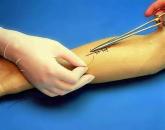


Popular
- Sinupret - complete instructions
- Number of antennae in arachnid and insect crustaceans
- Help for enterobiasis for the pool, how much is valid
- Ceftriaxone suspension. What is ceftriaxone?
- Third degree breast cancer: prognosis and treatment
- Salt Scrub for the scalp
- Do girls how many centimeters to the uterus
- Medications for the common cold: which are the most effective and safe?
- White mucous discharge after childbirth
- What is oral cancer, its profilatika, symptoms and treatment