Thick nipple. Why seals may appear. Bump on the nipple
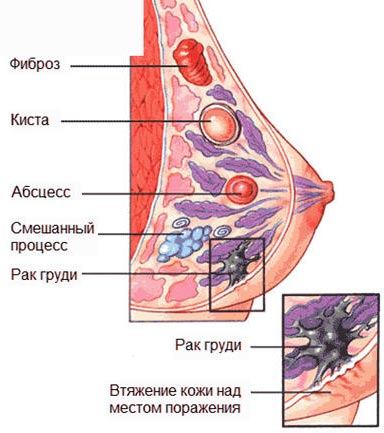
A lump in the mammary gland is a symptom of benign or malignant neoplasms of a nodular or diffuse form. Determining the causes, form and nature of the pathology helps to establish the need for treatment or careful monitoring of the patient's condition.
To exclude more serious diseases, it is necessary to conduct clinical studies of breast cancer.
Breast Inflammation Factors
A lump in the breast during lactation indicates a blockage of the lacrimal ducts caused by unstable or incomplete emptying. At elevated temperature, weakness and inflammation of the lining, a general blood test is prescribed to identify mastitis.
The formation of dense mobile areas before the menstrual cycles signal the manifestation of a benign tumor in the gland. She tends to change the location and parameters. Common pathologies are:
1. Advanced excretory duct.
2. Benign education.
3. Purulent inflammation of the tissues.
4. Blood clots.
5. Wen.
6. Local necrosis of breast fatty tissues.
For nodes characterized by discharge from the nipples. When detecting brownish shades, the risk of cancer increases. Hormonal imbalances, benign neoplasms and breast cancer are the main factors for the formation of seals in the gland.
Tumors on the background of pregnancy and feeding
Deformation of the glands under the influence of mammotropic, steroid and polypeptide hormones is observed throughout the entire period of pregnancy. At birth, hormonal production decreases. Under the influence of increased secretion of prolactin, milk is produced.
According to statistics, a large lump in the chest is observed in 3-4 thousand patients per year. The presence of a large cone is regarded as a natural phenomenon with hormonal changes. The disease is found in the latter stages. If you suspect a pathology, a comprehensive examination is appointed. A biopsy is performed under local anesthesia.

Fibrocystic disease
Mastopathy is a benign tumor of the breast. Symptoms of pathology are painful in nature, progressing a few days before the menstrual cycle. Inflamed areas grope independently.
Nodular mastopathy
The disease has the appearance of balls, clusters - cysts, the chest does not hurt. Characteristic features are serous or greenish discharge. The tumor is not soldered to the skin and halo. They are characterized by mobility, in the prone position cysts are not palpable. The lymph nodes located near are increased.
Diffuse neoplasm
Inflammation is characterized by a uniform increase in nodes and strands that disrupt the shape of the ducts and the lobes of the mammary glands. Small cysts that transform into focal seals or nodular proliferates are considered as a risk factor for oncology. Making a secret is diagnosed as adenous mastopathy. The proliferation of connective tissue signals the appearance of fibrous lesions in the mammary gland.
Mixed diffuse tumor
Puffiness, weighting and enlargement of the gland with aching pain 5-7 days before the onset of menstruation is regarded as a mixed type of diffuse neoplasm. The cause of the disease lies in the increased secretion of steroid sex hormones. The problem is exacerbated by late childbirth, abortion, disruptions in the menstrual cycle, thyroid inflammation or uterine trauma. Cases with late or early menopause are considered separately.
Benign lesions on the chest
Among common tumors, there are nodular and leaf-shaped types of fibroadenomas (tightly shaped capsules, with a densely elastic consistency). Sheet-like growth forms rapidly expanding layers in the gland. The skin becomes thinner, with a bluish tint. In 10% of cases, malignant degeneration is observed.
The formation of single nodes is observed in women under 35 years of age. Enlarged glands and connective tissues take a round shape with a clear outline and in size from 2 mm to 5 cm. The tumor is not painful. In patients aged 40 years, malignant transformations of the nodular adenofibroma are progressing.
![]()
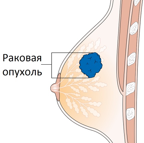
Breast cancer
The cause of cancer is an excess of steroid hormones (estrone, estriol, estradiol), contributing to the growth of the epithelium of the lobes and ducts, immature cells. Diagnostics allows to identify nodular and diffuse type of inflammation.
Nodal form of breast oncology
The dense knot with indistinct contours and a cover expands in the top direction, forming the wrinkled form of a nipple. This leads to his puffiness. Spreading through the ducts of the glands, the nipple is retracted and the cover is deformed. The cause is caused by swelling and an increase in skin follicles over inflammation.
Diffuse malignant neoplasm on the chest
Diffuse lump during pregnancy and feeding proceeds painlessly, but dramatically increases in size, negatively affecting the lymph nodes. Mastitis-like diffuse formation is diagnosed in adulthood. Brownish discharge accompanied by uneven puffiness and redness. Armored breast cancer affects size reduction and reduced mobility of the seal. The top cover has a shell shape.
Chest tumor tests and treatment
The mammologist's task is to identify the nature and factors in the neoplasm, confirmed by clinical studies:
x-ray;
mammography;
ductography;
Ultrasound;
laboratory tests (checking secretions).
An integrated approach to the survey allows you to establish the correct diagnosis and determine the method of treatment. To combat a diffuse tumor, conservative therapy is prescribed. Identifying nodes requires surgical intervention. An integrated approach aimed at treating oncology.
As practice shows, the bumps in the mammary gland are detected by a woman alone or by a gynecologist at the reception. However, the gynecologist does not make a diagnosis, he sends to a specialist in pathology of the female breast - mammologist.
Depending on the size of the cones, their localization, quantity, density and mobility, the mammologist can only make assumptions about the diagnosis, but accurate data allow us to obtain an instrumental examination and laboratory tests.
Types of tumors in the mammary gland
Depending on the nature of the bumps (seals, lump, knot), the following palpable mass can be distinguished:
- lipoma;
- fat necrosis;
- cyst;
- adenoma;
- lactostasis;
- calcinate;
- thrombosis.
It must be said that not each of these formations necessarily appears during self-examination. If the lump or seal is less than 10 mm in diameter and located on the periphery of the breast, it is difficult to detect it yourself. In such situations, tumor clusters are discovered by chance, in the course of other studies.
Methods for diagnosing tumors
To obtain the maximum possible amount of information about the parameters, character, location, behavior of a pathological cluster of cells, specialists use several instrumental methods of examination:
- chest x-ray (mammography);
- ductography (x-ray of the mammary gland with pre-inserted contrast into the ducts);
In addition, laboratory tests of punctate and lactate (cyst contents, discharge from the nipple) are used to confirm the status of the tumor and a biopsy is performed. It is worth recalling that, of all the formations found in the breast, only 5-10% are malignant. Often, laboratory tests are important for the exclusion of cancer, the possibility of choosing a conservative treatment and preserving the integrity of the breast.
Lipomas: causes, development, treatment
Tumor consolidation consisting of fat cells refers to lipomas. The causes of their occurrence in the mammary gland are not reliably identified, therefore, they most often include hormonal disorders, reduced immunity, nutritional problems, inflammatory or infectious processes, which take a long time in the body.
Lipoma in adipose tissue is formed due to uncontrolled division of cells of this type in one place, often in the sebaceous duct. Over time, education becomes independent, forming a bag around itself. Most often, breast lipomas are found in older women, less commonly in nursing mothers.
Treatment of small lipomas that do not bring discomfort is not indicated. If the fatty tumor interferes with the normal functioning of the breast, it is removed. Malignant lipomas occur extremely rarely, in less than 1% of cases.
Fat necrosis in the mammary gland
The death of the cells of adipose tissue often leads to their rotting, the formation of abscesses. To the touch fat necrosis is a soft movable seal, often inseparably associated with the skin. If the area around the nipple is affected, it can be retracted.
Instrumental diagnostics often do not give specific results, on a mammogram necrosis is similar to a cancerous tumor. Puncture and biopsy do not give results, since there are no tissues in the sample, only liquid. Sometimes the patient herself can assist in the diagnosis if she links the formation with a recent chest injury.
Treatment for fat necrosis is not carried out, only removal is recommended, since the picture of development is very similar to the malignant process.
In the study of the mammary glands, a number of seals of a different nature, formed in the glandular, stromal tissues or ducts, was called mastopathy. Mastopathies are characterized by different in shape, soreness, structure, seals, however, in 95% of cases are benign.

Cones of different nature and density are combined under the term "mastopathy" because of its hormone dependence. With a high degree of reliability, a link has been established between mastopathies and the predominance of estrogen over progesterone, excessive production of prolactin and secretion of the thyroid gland.
If the compaction is soft, with clear contours, but immobile relative to the gland, most likely it is a cyst. Large cysts that are detectable by touch pierce and eliminate fluid. The little ones are subjected to conservative treatment.
Fibrous formations are loose to the touch, but are often defined as seals. They consist of connective tissue in which atypical processes of cell division take place. Often, fibrous seals are noticeable before menstruation and in its first days, on days 5-6, spontaneously disappear. If treatment is required, emphasis is placed on anti-hormonal therapy.
Adenoma, fibroadenoma - a bump that hurts when it is palpated. Its contours are clear and smooth, the correct form. Such a formation, in contrast to cysts and fibrosis, refers to tumors, so a biopsy is often prescribed for accurate diagnosis. Such seals have an increased interest in diagnosis, often with large sizes, the doctor insists on removal. That adenomas are prone to malignancy. Small, isolated adenomas are observed, conservative treatment is used.
Lactostasis Cones
If you examine the photo of the breast during lactostasis, you can see a clear violation of the smoothness of the skin surface, redness, swelling. Lactostasis, with the exception of special cases, occurs in nursing mothers and is a blockage of the milk duct. The resulting plug does not allow milk to go freely through the duct, the gland lobe overflows, rough, forming a clearly expressed lump. Such a seal is noticeable not only to the touch, but also visually.
Many women are trying to pay great attention to their own breasts: beauty, size, etc. When chasing fashion trends and attractiveness, women forget to pay attention to their own health, as well as to a seal above their breasts. There are many diseases that cause condensation in the chest, as well as redness around the papillary area. Some illnesses may not remind of themselves at all until there is a serious threat to the patient’s health or life. Therefore, if women have any suspicious discomfort in the mammary gland or there is a seal over the breast, you should immediately contact a specialist in order to receive qualified treatment. Such checks need to pass from 22 years. Folk remedies can only be further treated, but only after consulting a doctor.
Why seals may appear
Condensation in the breast often occurs due to several factors:
- hormonal balance is impaired;
- the appearance of a benign neoplasm;
- development of breast cancer.
At 22, a woman should be regularly examined by a breast specialist, since from this age to 50 years, the risk of developing tumors increases. Many of the diseases causing tightness on the chest do not pose a serious threat, but it is not recommended to leave them unattended, as well as to conduct unauthorized intervention by folk remedies.
Important! Generalized swelling is often felt like a lot of small bulges (balls) that occur in the inside of the breast, on the left side, in the upper zone, near the nipple, etc. Also, there is excessive sensitivity.
Especially often these breast lumps appear before menstruation. New growths appear in any part of the mammary gland of women, disappear, change their volume in a matter of days, so there is no need to do anything to eliminate them.
Compaction over the breast before equated to pathologies, but today it is very common and is considered the norm. This phenomenon disappears after menopause, but can be detected in women who undergo hormone therapy after the onset of menopause.
Occurrence in pregnant women and during breastfeeding
In pregnant women, hormonal changes reach a global scale, which is an absolute norm. In a similar period of life, the breast begins to grow. The lactogenic effect of prolactin is suspended because of the high levels of progesterone and estrogen until the moment of delivery. After the birth has already begun, the concentration of the above hormones decreases sharply. Prolactin begins to be actively produced and as a result lactation occurs. Therefore, such breast lumps may be the norm.
Statistics claim that each of the 4 thousand women who have breast tightness, find a malignant neoplasm. During this period, it is much more difficult to detect it at the initial stage, because the seal inside the female breast can be attributed to hormonal changes. Such reasons affect the fact that a malignant tumor is determined in the later stages, when treatment is much more difficult and ineffective. Therefore, during this period, it should be carefully examined in the form of mammography, etc. The same applies to women in the postpartum period. Any redness on the right or left breast, breast lump requires a qualified examination in order to provide treatment in time.
Compaction with a tumor of benign nature
Condensation in the mammary gland may indicate the presence of a benign tumor - fibroadenoma. A similar disease can occur in women at the age of 22, and its shape resembles a single nodule. The structure of the tumor includes connective and sinewy tissue. It has clear edges, has a rounded shape and does not exceed 5 cm in size. These signs may be the only ones that indicate the presence of the disease, as it can proceed without any pain. Such a breast seal may not require special medical intervention and go by itself. Treatment conducted by folk remedies, can only aggravate the situation.
Cancer Seal
Breast induction may be one of the symptoms that indicate breast cancer. The causes of the disease may indicate an increased estrogen content in the body of women. After the increase of this hormone, the lobules of the epithelium and ducts may begin to expand, which affects the formation of atypical cells. Such seals on the chest are a threat to the life of the patient. In the initial stages, the tumor is very difficult to detect. It is found by chance on scheduled checks or after self-examination. In the later stages, you can feel the characteristic condensation in the armpit, as the tumor begins to grow.
There are two forms of cancer:
- Diffuse
- Nodal.
The second is more common. The reasons for its appearance indicate a hormonal imbalance. It represents a tight knot, which does not cause any pain. Breast compaction has blurred outlines and a rough surface. The neoplasm usually grows in the direction of the skin, which affects its external characteristics. The skin near the affected area becomes retracted, wrinkled. After that, the seal on the chest begins to be accompanied by swelling near the areolas and nipple. Orange peel skin is swollen with enlarged follicles over the affected area. It is very difficult to cure the disease if it began to show all its symptoms. Usually, between the ages of 22 and 32, a tight seal may occur in women,  who live in an ecologically polluted area.
Diffuse form of the tumor appears in women after childbirth, during pregnancy or breastfeeding. Causes lie also in hormonal jumps. The neoplasm may not give any signs and pains for a long time. However, it quickly grows and metastasizes to nearby lymph nodes. Girls at 22 may also have this disease. Therefore, it is advisable to undergo appropriate examinations after childbirth, during pregnancy and during lactation in order to start treatment in time. Intervention folk remedies of any type must be agreed with the doctor.
Important! Any compression in the mammary gland requires the attention of a qualified breast specialist. Only after palpation and diagnostic measures can one determine whether a seal above the breast is a threat to the patient’s life and what treatment should be prescribed.
What symptoms should pay attention
The reasons for the development of a tumor in the breast are different. But there are a number of symptoms, in addition to the presence of a lump in the chest, which should cause women to be wary and after their detection, they are obliged to consult a doctor:

Similar signs can appear in girls at the age of 22, therefore it is necessary to do something. To detect this pathology, you need to conduct regular self-examination.
The presence of blood clots
 Consolidation under the breast or on it in women can be a sign indicating a blood clot in the veins. The following symptoms are associated with this phenomenon:
- Swelling near the passage of the vein.
- Pain sensations.
- Local temperature rise.
- Change in skin tone.
This phenomenon is quite rare, but breast tightening in women may indicate its presence.
In order to start treatment on time and get rid of the disease, you should regularly visit the doctor, because the disease of the mammary glands can also manifest itself in young years. Therapy with folk remedies can also be used, but if allowed by experts.
Important! In order to achieve recovery, it is necessary to prescribe a qualified treatment after the patient undergoes a series of diagnostic studies. Folk remedies can not be cured of diseases of the mammary glands. They can be used as adjuvant therapy, after consulting with a doctor.
Lump in chest - an alarming symptom that worries many women. Under the mask of a small seal in the breast can hide various diseases. Some of them do not pose any danger to the life and health of women, while others require close attention and an urgent appeal to a specialist.
Before a visit to the doctor, it is necessary to clearly understand where the seal is located (in the chest, on the nipple, under the armpit), and also to assess the presence or absence of other symptoms. This can play an important role in making an accurate diagnosis and choosing the right treatment.
It is very suspicious if there is a lump in the chest, that is, directly in the thickness of the mammary gland. It can be easily identified by breast self-examination. It is necessary to evaluate not only the size of the seal, but also the degree of its mobility, whether it is not spliced with the surrounding tissues. On this depends the accuracy of diagnosis and subsequent therapeutic tactics.
Fibrocystic mastopathy
One of the most common diseases in which a lump occurs in the thickness of the mammary gland is mastopathy, namely its fibrocystic form. The development of this disease is caused by hormonal disorders, as a result of which individual parts of the mammary gland overgrow with the formation of limited cavities (cysts) and nodes.
As a rule, the process affects both mammary glands, although in one of them the changes may be more pronounced. In addition to the nodes and seals women complain of discomfort and pain in the chest, changing during the menstrual cycle, as well as unusual discharge from the nipple. Some women have small seals under the arm.
Although the symptoms of mastopathy look quite threatening, it is a completely benign process that does not go into cancer. For treatment, hormone therapy is used, in extremely rare cases, removal of nodes is required.
Benign breast tumor
Often the lump in the breast is a benign tumor of the breast. As a rule, this is a single formation, moving relative to the surrounding tissues.
Benign breast tumors differ in their microscopic (histological) structure. The most common neoplasms are:
- Fibroadenoma;
- Intraductal papilloma;
- Lipoma;
- Breast cyst.
Some of these tumors arise as part of mastopathy, while others develop independently. The reasons for their appearance are not fully understood, it is assumed the role of hereditary factors, lifestyle and adverse environmental conditions.
Treat benign breast tumors only operatively. Conservative and traditional methods of treatment are ineffective.
Breast abscess
This is not a common disease, occurring mainly in women who are breastfeeding. As a rule, an abscess, or a limited area of purulent fusion, develops on the background of mastitis - inflammation of the breast. This disease occurs as a result of the ingestion of pathogenic bacteria into the gland ducts, as well as in violation of milk outflow (for example, during its stagnation).
Breast abscess
Breast abscess - This is a round, sharply painful formation in the thickness of the chest, accompanied by severe throbbing pain. Other symptoms of inflammation are also characteristic, such as:
Redness of the affected breast;
- Purulent discharge from the nipple;
- Swollen lymph nodes under the arm;
- Increased body temperature;
- General malaise and weakness.
A woman who has been diagnosed with an abscess of the mammary gland should immediately stop breastfeeding. Appointed a powerful antibacterial and detoxification therapy, which is designed to destroy the bacteria. An abscess is usually treated surgically, but in some cases, conservative therapy can achieve complete recovery.
Mammary cancer
The most disturbing reason for the appearance of a lump in the breast is breast cancer. The cunning of this disease is that it is asymptomatic for a long time. The woman whose breast cancer was detected at the first stage was lucky, because in this case you can achieve a 100% recovery. However, as the process progresses and the tumor grows, the chances of not only healing but also survival are rapidly decreasing.
Breast cancer is defined as a dense tumor that is adherent to the surrounding tissues. In the later stages, nipple retraction is characteristic, as well as a change in the appearance of the breast - the skin takes on the appearance of a “lemon peel”, or even ulcerates. Lymph nodes under the armpits are enlarged, sometimes they are much larger than the primary node in the chest. This is due to the metastasis of the tumor and the progression of the process.
Breast cancer treatment is a complex task that a woman solves with an oncologist. Usually, complex surgical, chemotherapeutic and radiation treatments are used. The volume of the operation depends on the size of the tumor, the degree of germination in the surrounding tissues, as well as the presence of metastases. In the first stage of cancer, when the primary focus is still quite small, a simple excision with breast preservation is sufficient. In advanced cases, amputation of the mammary gland, removal of the pectoralis major muscle and lymph nodes under the armpits are required.
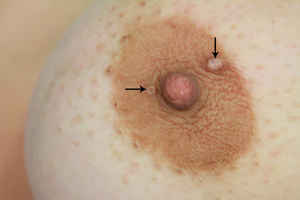
Bump on the nipple
Another problem with which women refer to a mammologist is a bump on the nipple or in the areola. As a rule, these are benign neoplasms that cause only aesthetic inconvenience. However, in some cases, growth on the nipple may be a manifestation of a malignant process.
Most often on the nipple occurs papilloma (wart). This is a dense formation resembling a cauliflower. The cause of papilloma is HPV - human papillomavirus. Treatment consists of surgical removal of the tumor.
Small whitish seals in the areola area of the nipples are Montgomery's tubercles. According to their structure, they are secreted glands. When the duct is blocked, the secret accumulates in the lumen of the gland, causing its enlargement and inflammation. This problem does not pose a danger to the life and health of women, delivering exclusively aesthetic inconveniences.
Seal under the arm
Finally, some women are worried about the appearance of a bump or seal under the arm. It should be remembered that in many people the lymph nodes under the armpit normally reach a size of up to 2.5 cm, which is completely normal. There are many reasons for their increase, and they concern not only the mammary gland. In particular, this is possible with any infectious disease, blood diseases and so on.
However, sometimes an enlarged axillary lymph node may be the first sign of an oncological process in the mammary gland. To exclude this condition, it is necessary to consult a specialist.


Popular
- Blood disorders in children
- What can not drink a pill
- Blood disorders in children
- Blood disorders in children
- Bones: structure, composition, types of bones, types of connections and their characteristics
- How many live with lung cancer
- Influenza in children: how to treat, what can and cannot be done to parents, what medicines will help?
- "They are people too ..." - you say
- What helps "Prednisolone"
- How are menstruation for endometriosis?