The structure of the middle ear. Features of the structure of the semicircular canals. The structure of the middle ear of a person
The ear is a complex organ of humans and animals, thanks to which the sound vibrations are perceived and transferred to the main nerve center of the brain. Also, the ear performs the function of maintaining balance.
As everyone knows, the human ear is a paired organ, located in the thickness of the temporal bone of the skull. Outside, the ear is bounded by the auricle. It is the direct receiver and conductor of all sounds.
Operation is good to hear
A small scar for the new ear. Placing bone prostheses is often practiced in children aged 7 to 8 years old, when the origin of hearing problems can be clearly identified. If the child continues to grow, it is believed that in 7-8 years the bones have reached their final size. Only ear cavities will grow, but this will not change the function of the prosthesis.
Inner ear - This is the part of the ear that contains the organ of hearing, but also the organ of balance. In the inner ear there are two different entities: it is a cavity in which all sensory structures are located inner ear and located inside the bone labyrinth. When we talk about the inner ear, we are talking about a liquid medium, and the entire inner ear is embedded in a liquid called perilymph. The membrane labyrinth is built into the perilymph, which separates it from the bone labyrinth. In addition, the membrane labyrinth contains another fluid, called the endolymph.
The hearing aid of a person can perceive sound vibrations, the frequency of which exceeds 16 Hertz. The maximum threshold of ear sensitivity is 20,000 Hz.
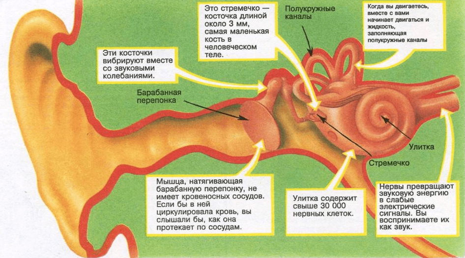
Human ear structure
The composition of the human hearing aid includes:
- The outer part
- middle part
- Interior
In order to understand the functions performed by one or another constituent parts, you need to know the structure of each of them. Rather complex mechanisms of transmission of sounds allow a person to hear sounds in the form in which they come from outside.
Bone Maze contains
The lobby, the cavity that opens into the drum box through an oval window, the latter being blocked by a plate. Semicircular canals, which are canals in the form of semicircles and connected to the lobby. Bone snail, which is a wound spiral wound and cut in half along the entire length, except for the upper part of the blade, called the spiral blade. Thus, two ramps of a bony snail are formed, one of which opens in the lobby, this is the vestibular ramp, the other, which opens in a round window, is a drum ramp. Membrane labyrinth is a bag with a very thin wall, it contains the utrikl and the bag contained in the run-up, the organ that plays a role in balance, we will not deepen this moment, because it does not fit into our problematic, it also contains membrane semicircular channels contained in the semicircular bone canals, it eventually contains a membrane cochlea.

- Inner ear. Is the most difficult part of hearing aid. The anatomy of the inner ear is quite complicated, so it is often called the labyrinth labyrinth. It is also located in the temporal bone, or rather, in its stony part.
The inner ear is connected to the middle one by means of oval and round windows. The webbed labyrinth consists of a vestibule, a cochlea and semicircular canals filled with two types of fluid: the endolymph and the perilymph. Also in the inner ear is the vestibular system responsible for the balance of a person, and his ability to accelerate in space. Oscillations that have arisen in the oval window, go to the liquid. With it, irritated receptors that are in the cochlea, which leads to the formation of nerve impulses.
The vestibular apparatus contains receptors that are located on the crista of the canals. They are of two types: in the form of a cylinder and a flask. The hairs are opposite each other. Stereocilia during excitation cause excitement, and kinocilium, on the contrary, contribute to inhibition.
The snail is an organ of hearing. The cochlear nerve is located in the cavity of the columella: the axis around which the snail is wound. The snail consists of three ramps: a vestibular ramp, a cochlear ramp or a cochlear canal and a drum ramp. A spiral blade separates the vestibular ramp from the tower ramp. They belong to the bone maze, so they contain perilymph, and they communicate over their limbs, the drum ram opens on a round window and the vestibular ramp in the lobby.
It is this complexity in the anatomy of the inner ear that can allow the movement of the fluid of the latter. The cochlear ramp is part of the membrane labyrinth, it is located between the vestibular ramp and the tympanic ramp, glued to the vestibular ramp and contains the endolymph. Reissner's membrane is a membrane that separates the cochlear canal from the vestibular ramp, and the basilar membrane will serve as a support for the organ of cortical, this is the second side of the cochlear ramp. This membrane plays an important role because it has a frequency of vibration characteristic of all points, so the inner ear is able to recognize the frequency of the sound.
For a more accurate understanding of the topic, we offer you a photo diagram of the structure of the human ear, which presents the complete anatomy of the human ear:
As you can see, the human hearing aid is a rather complex system of various formations that perform a number of important, irreplaceable functions. With regard to the structure of the outer part of the ear, then each person may have individual characteristics that do not harm the main function.
Sharp sounds predominantly distort the base of the membrane, bass sounds, the top. In this diagram, the base membrane unfolds. The cochlear canal essentially contains the organ of Corti. It is in this organ that we will find the ciliary auditory cells that are in contact with their base, with the nerve fibers, while their end is in contact with the tectorial membrane. This membrane is a fibrous membrane that covers the entire body of Corti. The organ of Corti is supported by two large cells called the columns of Corti.
Care of the hearing aid is an integral part of human hygiene, since hearing loss and other diseases associated with the outer, middle or inner ear are possible as a result of functional impairment.
According to research scientists, a person is more difficult to tolerate the loss of vision than the loss of hearing, because it loses the ability to communicate with the environment, that is, it becomes isolated.
This set of pillars is called the Arcade of Corti. On both sides of this arch there are internal and external hair cells. The spiral ganglion is a channel containing nerve cells. These cells spread to the nerve fibers in contact with the hair cells. Thus, the hair cells are in contact with the auditory nerve.
The snail is a part of the inner ear that interferes with the hearing, so this is a place of mechanical phenomena, but also electrical. Due to the complexity of the anatomy of the inner ear, fluid movement can occur. Earlier we saw that the drum and vestibular ramps contain perilymph, communicate with each other on the top of the cochlea, and open up accordingly to a round window and an oval window. Thus, when the stirrup plate sinks into the oval window, the round window is rounded out due to the liquid pushed by the plate.
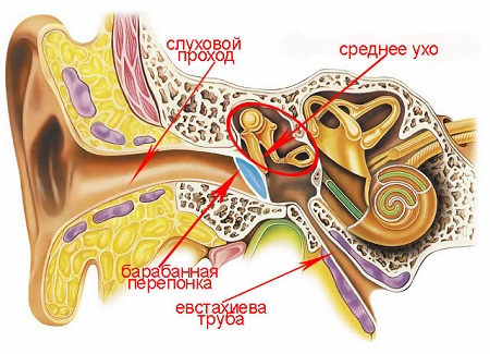
The middle ear (auris media) consists of several interconnected air cavities: the tympanic cavity (cavum tympani), auditory tube (tuba auditiva), the entrance to the cave (aditus ad antrum), the cave (antrum) and the air cells associated with it mastoid (cellulae mastoidea). Through the auditory tube, the middle ear communicates with the nasopharynx; under normal conditions, this is the only message of all cavities of the middle ear with the external environment.
When movements occur in the inner ear, all structures of the cochlear ramp move together. Thus, when the vibrations are transmitted by the rod, we observe the vibrations of this set, also called: the wall of the cochlea. The frequency of vibration is maintained due to the basilar membrane, which will allow to recognize the frequency of sound. As in all organs, the functioning of the inner ear is associated with electrical activity, therefore, when the sound vibrations reach the inner ear, this is the place of changes in electrical potential: the cochlear microphone potential and the potential of summation.
Fig. 4.4.
1 - horizontal semicircular canal; 2 - channel facial nerve; 3 - the roof of the tympanum; 4 - window of the vestibule; 5 - muscle canal; 6 - tympanic hole of the auditory tube; 7 - carotid canal; 8 - promontorium; 9 - drum nerve; 10 - jugular fossa; 11 - window of the cochlea; 12 - drum string; 13 - pyramidal process; 14 - cave entrance.
Outer ear. It consists of the auricle and the external auditory canal. Auricle have a cartilaginous structure and an irregular lateral surface due to the presence of multiple folds, it collects sounds and transmits them to the outer auditory canal. This is a cylindrical bone-like canal in the medial and cartilaginous third in the lateral two-thirds, it is directed from the outside to the inside and from the front to the back and ends with the eardrum, a complex fibrous structure that separates the outer ear from the middle ear.
In this channel there are some glands that secrete an ear wax, which performs the function of protecting the auditory canal from the entry of foreign substances. Middle ear. This cavity is filled with air, dug into a temporary bone, and it is so thin that it can barely contain 5-6 drops of water. This cavity contains three bones of hearing: a hammer, an anvil and a stirrup, which are connected together and in the drum box with the help of joints and ligaments. They form a kind of chain that goes from the tympanum membrane to the oval window.
Barbarna N p I l about with about t (Fig. 4.4). Drum cavity can be compared with a cube of irregular shape up to 1 cm3. It distinguishes six walls: upper, lower, front, back, outer and inner.
At the top of the frame, or the roof, the tympanic cavity (tegmen tympani) is represented by a bone plate 1-6 mm thick. It separates the tympanic cavity from the middle cranial fossa. In the roof there are small openings through which the vessels pass, carrying blood from the dura to the mucous membrane of the middle ear. Sometimes in the upper wall of de-formation; in these cases, the mucous membrane of the tympanic cavity is directly adjacent to the dura mater.
In the hammer, you can distinguish between a pen and a head. The handle is attached to the inner surface of the eardrum, and the head is circularly articulated with the anvil. The latter is attached on one side to the hammer, and the other to the bracket, which in turn rests on the membrane of the oval window.
In front, the middle ear cavity is connected to rhinos, that is, that part of the pharynx, located behind the nasal cavities, through the Eustachian tube. Thanks to this channel, the pressure exerted on the inner surface of the membrane of the eardrum is equal to that which exerts on the outer surface, that is, the atmospheric pressure. When swallowed or gaped, the air quickly passes through the opening of the tube, which makes it possible to compensate for the decrease in pressure inside the eardrum due to the continuous absorption of air associated with the physiological processes of gas exchange with extracellular fluids.
In infants and children of the first years of life, an unpainted gap (fissura petrosquamosa) is located on the border between the pyramid and the scales of the temporal bone, causing their cerebral symptoms in acute inflammation middle ear. Subsequently, a suture is formed at this place (sutura petrosquamosa) and communication with the cranial cavity is eliminated at this place.
This mechanism is of fundamental importance, since the tympanic membrane can optimally vibrate only when the pressures on its sides are equal. It consists of a series of cavities dug into the temporal bone, called the bone labyrinth, which contain membranous structures that repeat their shape, called the membrane labyrinth. The cavities that make up the bone labyrinth are a vestibule, a snail, and three semicircular canals. The membrane labyrinth is formed by the utricle and sacculus contained in the vestibule, the cochlear canal inside the cochlea and the semicircular membrane channels inside the bone.
N and I (I remn I) with tehnka, or the bottom of the tympanic cavity (paries jugularis), borders on the jugular fossa (fossa jugularis) lying below it, in which the jugular vein is located (bulbus venae jugularis). The larger the fossa juts into the tympanum, the thinner the bony wall. The bottom wall can be very thin or have de-ejections, through which the bulb of the vein sometimes bulges into the tympanic cavity. This leads to the possibility of injuring the bulb of the jugular vein, accompanied by severe bleeding, during paracentesis or careless scraping of granulations from the bottom of the tympanic cavity.
The lobby is located between the cochlea and the semicircular canals and communicates with the middle ear through an oval window. Utrikl and the bag contain on their walls cells equipped with eyelashes, on which a membrane is placed, inside which there are small crystals of calcium carbonate, called otoliths. The otoliths make the membrane sensitive to gravity, and then bend it differently when the body changes position, causing eyelash flexion. Sensory cells are associated with nerve fibers that belong to the vestibular nerve.
T e c h i n, a tubular or carotid (paries tubaria, s.caroticus), the tympanic cavity is formed by a thin bone plate, outside of which is located the internal carotid artery. In the front wall there are two openings, the upper of which, narrow, leads to the canal for the muscle that pulls eardrum (semicanalis m.tensoris tympani), and the lower, wide, - in the tympanic mouth of the auditory tube (ostium tympanicum tybae auditivae). In addition, the front wall is penetrated by thin canaliculi (canaliculi caroticotympanici), through which vessels and nerves pass into the tympanic cavity, in some cases it has dehydration.
The movement of the head causes contact between the membrane and the cilia, causing activation of the ciliary cell, which stimulates the nerve receptors, so that the nerve conducts impulses to the brain, where sensations associated with gravity are processed. reflection straightening. The snail forms the front part of the bone maze, resembles the snail shell and consists of a spiral channel that runs about two and a half turns around the central conical cone. Inside there is a cochlear canal, which divides the inner space of the cochlea into two parts, one on top and one below the cochlear canal itself.
Za d ia i (io s cc e c d ia d ia) c ta n a and the tympanic cavity (paries mastoideus) borders on the mastoid process. In the upper section of this wall there is a wide passage (aditus adantrum), which reports a dimple - attic (attic) with a permanent cell of the mastoid process - a cave (antrum mastoideum). Below this course is the bone protrusion - the pyramidal process, from which the stapedius muscle begins (m.stapedius). On the outer surface of the pyramidal process there is a drum opening (apertura tympanica canaliculi chordae) through which the drum string (chorda tympani) extends from the facial nerve into the drum cavity. In the thickness of the lower section back wall passes the descending knee of the facial nerve canal.
Inside the cochlear duct, the organ of Corti is maintained throughout its length. It consists of supporting cells and acoustically sensitive cells that represent the auditory receptors. They are equipped with eyelashes that get wet in the fluid of the cochlear canal. A tectoral membrane is placed above the organ of Corti, which overlaps the hair of the acoustic cells. When the base of the cochlear canal vibrates, it causes the hair to approach the tectoral membrane, which causes activation of the acoustic cells.
The state of excitation is transmitted to the acoustic centers of the brain through the nerve fibers of the cochlear acoustic nerve. There are three semicircular bone canals, inside of which there are membrane semicircular canals. Each of them contains a liquid and is associated with the uterus, which is located in the bone vestibule. One of the two limbs of each canal expands in the form of an ampoule, inside these dilatations there are ciliary cells in which gelatinous substance that does not contain otoliths predominates. With rapid movements of rotation of the head and body, endolymph flows are formed, which flex eyelashes, causing excitation of nerve cells, and activation of reflexes, which tend to keep the body in balance and vision stable.
The nasal ligament (n e p a n t a t ya) with the tympanic cavity (paries membranaceus) is formed by the eardrum and partly in the region of the attic by the bone plate, which extends from the upper bone walls of the external auditory canal.
V n u t r e n i (l a b and r and n t n i, med and a l n i, pro ra m o n t o r and a l n i am ) The tympanic cavity (paries labyrinthicus) is the outer wall of the labyrinth and separates it from the cavity of the middle ear. In the middle part of this wall there is an elevation of an oval shape - a promontory (promontorium), formed by the projection of the main curl of the cochlea.
To the rear and to the top of the cape there is a niche of the vestibule window (oval window according to the old nomenclature; fenestra vestibuli), closed by the base of the stirrup (basis stapedis). The latter is attached to the edges of the window by means of an annular ligament (lig. Annulare). In the direction backwards and downwards from the cape there is another niche, at the bottom of which there is a cochlea window (a round window according to the old nomenclature; fenestra cochleae), leading to the cochlea and closed by the secondary eardrum (membrana ympany secundaria), which consists of three layers: outer - mucous, middle - connective tissue and internal - endothelial.
Above the window of the vestibule, along the inner wall of the tympanic cavity in the direction from front to back passes the horizontal knee of the bone canal of the facial nerve, which, reaching the protrusion of the horizontal semicircular canal on the inner wall of the antrum, turns the vertically downward knee down and out to the base of the skull through the stylospinal cavity (for. stylomastoideum). The facial nerve is located in the bone canal (canalis Fallopii). A horizontal segment of the facial nerve canal above the vestibule protrudes into the tympanic cavity in the form of a bone ridge (prominentia canalis facialis). Here it has a very thin wall, in which there are often devisations, which contributes to the spread of inflammation from the middle ear to the nerve and the occurrence of facial nerve paralysis. An otolaryngologist is sometimes faced with various options and anomalies of the location of the facial nerve, both in its tympanic and mastoid regions.
On the middle floor of the tympanic cavity, the tympanic string (chorda tympani) departs from the facial nerve. It passes between the malleus and the incus through the entire tympanic cavity near the eardrum and exits through the stony-tympanic (glazeru) slit (fissura petrotympanica, s.Glaseri), giving the taste fibers to the tongue on its side, secretory fibers to the salivary gland and fiber to the nerve vascular plexus.
The drum cavity is conditionally divided into three sections, or floors: the upper one is the attic, or the epitympanum, which is located above the upper edge of the stretched part of the eardrum, and the height of the attic varies from 3 to 6 mm. The articulation of the malleus with the incus in it divides the attic into the outer and inner sections. The lower part of the outer part of the attic is called the “upper recess of the eardrum,” or “Prussian space,” the posterior attic passes into the antrum; medium - the largest in size (mesotympanum), corresponds to the location of the stretched part of the eardrum; the lower one (hypotympanum) is a depression below the level of attachment of the eardrum (Fig. 4.5, a, b).

A - sagittal section: 1 - superior ligament of the incus; 2 - short leg of the anvil; 3 - cave; 4 - posterior ligament of the incus; 5 - long leg of the anvil; 6 - posterior hammer fold; 7 - back pocket of membrane; 8 - lenticular process of the incus; 9 - hammer handle; 10 - the facial nerve canal; 11 - barb string; 12 - facial nerve; 13 - drum ring; 14 - stretched part of the eardrum; 15 auditory tube; 16 — front hammer fold, 17 — front membrane pocket; 18 - ; 19 - hammer head; 20 - upper ligament of the malleus; 21 - anvil-and-articulation joint.
The mucous membrane of the tympanic cavity is a continuation of the mucous membrane of the nasopharynx (through the auditory tube); it covers the walls of the tympanic cavity, auditory ossicles and their bundles, forming a series of folds and pockets. Tightly adhering to the bone walls, the mucous membrane is for them simultaneously a periosteum (mucoperioste). It is covered mostly flat epithelium, with the exception of the mouth of the auditory tube,

Fig. 4.5. Continued.
: 22 - anterior semicircular canal; 23 - posterior semicircular canal; 24 - lateral semicircular canal; 25 - stapedial tendon tendon; 26 - VIII cranial (pre-door-cochlear) nerve; 27 - probe in the cochlea window; 28 - snail; 29 - muscle straining the eardrum; 30 - sleepy channel; 31 - stirrup; 32 - the anterior process of the malleus; 33 - the upper pocket of the eardrum (Prussac space); 34 - lateral ligament of the malleus.
Where there is a ciliated cylindrical epithelium. In some places of the mucous membrane there are glands.
The hammer and the malleus (malleus), the anvil (incus) and the stirrup (stapes) are connected by joints, anatomically and functionally represent a single chain (Fig. 4.6), which stretches from the eardrum to the window of the vestibule. The handle of the malleus is interwoven into the fibrous layer of the eardrum, the base of the stirrup is reinforced in the niche of the vestibule window. The main mass of the auditory ossicles - the head and neck of the malleus, the body of the anvil - is in the rebar space (see. Fig. 4.5, b). The auditory ossicles are reinforced between themselves and with the walls of the tympanic cavity with the help of elastic ligaments, which ensures their free movement during the movement of the eardrum.

1 - the anvil; 2 - long leg of the anvil; 3 - anvil-temporal joint; 4 - stirrup; 5 - rear leg of the stirrup; 6 - stirrup base; 7- front leg stirrup; 8 - hammer handle; 9 - the anterior process of the malleus; 10 - hammer; 11 - hammer head; 12 - anvil-and-articulation; 13 - the short process of the incus; 14 - the body of the anvil.
In the hammer there is a handle, neck and head. At the base of the handle is a short process, protruding outwardly part of the eardrum. The mass of the hammer is about 30 mg.
The incus consists of a body, a short appendix, and a long appendix articulated with a stirrup. The mass of the anvil is about 27 mg.
In the stirrup there is a head, two legs and a base.
The annular ligament, by means of which the base of the stapes is attached to the edge of the vestibule window, is quite elastic and provides good vibrational mobility of the stapes. In the anterior section, this ligament is wider than in the posterior, therefore, when transmitting sound vibrations, the base of the stirrup shifts mainly by its anterior pole.
The stirrup is the smallest of the auditory ossicles; its mass is about 2.5 mg with a base area of 3-3.5 mm2.
The braces are represented by two muscles: the tensioning tympanic membrane (m.tensor tympani) and the stapedius (m. stapedius). Both of these muscles, on the one hand, keep the auditory ossicles in a certain position, the most favorable for the sound, on the other - protect the inner ear from excessive sound stimulation by reflex contraction. The muscle tightening the eardrum is attached at one end in the region of the opening of the auditory tube, the other to the handle of the malleus near the neck. It is innervated by the mandibular branch. trigeminal nerve through the ear ganglion; the stapedial muscle starts from the pyramidal protrusion and is attached to the stirrup neck; innervated by the stapedal nerve (n.stapedius) with a branch of the facial nerve.
As it was already noted, the cavity (e in s t and x and e in a) is the formation through which the tympanic cavity communicates with the external environment: it opens in the region of the nasopharynx. The auditory tube consists of two parts: the short bone - 1L canal (pars ossea) and the long cartilaginous - 2/3 (pars cartilaginea). Its length in adults is on average 3.5 cm, in newborns it is 2 cm.
At the point of transition of the cartilaginous part to the bone, an isthmus (isthmus) is formed - the narrowest point (diameter 1-1.5 mm); it is located approximately 24 mm from the pharyngeal opening of the tube. The lumen of the bony part of the auditory tube in the section is a semblance of a triangle, and in the membranous-cartilage region the walls of the tube are adjacent to each other.
The internal carotid artery runs medial to the bone part of the tube. It should be noted that in the membranous-cartilaginous part of the lower and anterior walls of the pipe are represented only by fibrous tissue. The pharyngeal opening of the auditory tube is 2 times wider than the tympanic one and located 1-2.5 cm below it on the side wall of the nasopharynx at the level of the posterior end of the inferior nasal concha.
The cavity of the tympanic cavity is carried out from the basins of the external and partially internal carotid arteries: the anterior, the tympanic artery extending from the maxillary; posterior ear artery extending from the stylo-mastoid artery and anastomosing with the middle meningeal artery. From the internal carotid artery branches depart to the anterior sections of the tympanic cavity.
In the case of the tympanum from the tympanic cavity occurs mainly on the same veins.
L and m f about o Tt about to from the tympanic cavity should be along the mucous membrane of the auditory tube in the pharyngeal lymph nodes.
In addition, the tympanic cavity occurs due to the tympanic nerve (n.tympanicus) of the ninth pair (n.glossopharyngeus) of the cranial nerves. Having entered the tympanum, the tympanic nerve and its branches anastomose on the inner wall with the branches of the facial nerve, the trigeminal and sympathetic plexuses of the internal carotid artery, forming the tympanic plexus (plexus tympanicus s.Jacobsoni).
С о с цthе в и д Ы Ы о д р о (prosessus mastoideus).
In the newborn, the mastoid portion of the middle ear has the appearance of a small elevation behind the upper-rear edge of the drum ring, containing only one cavity — the antrum (cave). Starting from the 2nd year, this elevation is pulled downward due to the development of the muscles of the neck and neck. The formation of the process ends mostly by the end of the 6th - the beginning of the 7th year of life.
The mastoid process of an adult resembles a cone, tilted down by its top, a protrusion. Internal structure the mastoid process is unequal and depends mainly on the formation of pneumatic cavities. This process occurs by replacing the bone marrow tissue with the growing epithelium. As bone grows, the number of air cells increases. By the nature of pneumatisation, it is necessary to distinguish: 1) a type of structure of the mastoid process, when the number of air-cells is large enough. They fill almost the entire process and sometimes even extend to the scales of the temporal bone, the pyramid, the bony part of the auditory tube, the zygomatic process; 2) d and pl about e and t and with to and d (spongiosal, spongy) type of structure. In this case, the number of air cells is small, they look like small cavities limited by trabeculae, and are located mainly near the cave; 3) c kleorotichi and compact (compact) type of structure: the mastoid process consists of extremely dense bone tissue.
If the pneumatic type of the structure of the mastoid process is observed during normal development of the child, then diplomatic and sclerotic are sometimes the result of metabolic disturbances or the result of general and local inflammatory diseases etc. There is an opinion that some genetic or constitutional factors, as well as resistance and organotissue reactivity associated with them, have a definite influence on the process of pneumatization of the mastoid process.
The anatomical structure of the mastoid process is such that all of its air cells, regardless of their distribution and location, communicate with each other and with the cave, which through aditus ad antrum communicates with the drummed cavity. The cave is the only congenital air-cavity, its development does not depend on the type of structure of the mastoid process.
In infants, unlike adults, it is significantly larger in volume and is located quite close to the outer surface. In adults, the cave lies at a depth of 2-2.5 cm from the outer surface of the mastoid process. The size of the mastoid process in adults ranges from 9 to 15 mm in length, 5 to 8 mm in width, and 4 to 18 mm in height. In a newborn, the dimensions of the cave are the same. A cave separates from the dura mater of the middle cranial fossa (tegmen antri), if it is destroyed by a purulent process, inflammation can pass to the meninges.
The dura mater of the posterior cranial fossa is separated from the cavity of the mastoid process by the Trautmann triangle, which is located posteriorly from the facial nerve to the sigmoid sinus. The mucous membrane lining the cave and air cells, is a continuation of the mucous membrane of the tympanic cavity.
On the inner rear surface (from the side of the cranial cavity) of the mastoid process, there is a recess in the form of a groove. It contains the sigmoid venous sinus (sinus sigmoideus), through which venous blood flows from the brain to the jugular vein system. The dura mater of the posterior cranial fossa is delimited from the cellular system of the mastoid by means of a thin, but rather dense bone plate (lamina vitrea). In some cases purulent inflammation cells can lead to the destruction of this plate and the penetration of infection into the venous sinus. Sometimes a mastoid injury can cause a violation of the integrity of the sinus wall and lead to life-threatening bleeding. Near the cells of the mastoid process is the mastoid facial nerve. This neighborhood sometimes explains the paralysis and paresis of the facial nerve in acute and chronic inflammation of the middle ear.
Outside, the mastoid process has a compact osteo-cortical layer, the surface of which is rough, especially in the lower part, where the sternocleidomastoid muscle (m.sternocleidomastoideus) is attached. There is a deep furrow (incisura mastoidea) on the inner side of the process apex, where the digastric muscle (m.digastricus) is attached. Through this furrow, pus sometimes breaks out of the process cells under the neck muscles. Within the outer surface of the mastoid process is a smooth triangular platform, called the "Shipot triangle". In the front-top corner of this triangle are a hole in the form of a platform (planum mastoidea) and a scallop (spina suprameatum), which correspond to the outer wall of the antrum. In this area, bone trepanation is performed in search of a cave for mastoiditis in adults and antritis in children.
The cancer of the mastoid region is made from the posterior aural artery (a.auricularis posterior - a branch of the external carotid artery - a.carotis externa). The venture occurs in the vein of the same name, which flows into the external jugular vein (v.jugularis externa).
The nasal cavity of the mastoid area is provided by the sensory nerves from the upper cervical plexus of the large auricular (n.auricularis magnus) and the small occipital (noscipitalis minor). The motor nerve for the rudimentary ear muscles (m.auricularis posterior) is the same branch of the facial nerve.


Popular
- Breast cancer is curable at any stage.
- The remedy for the cold Sinupret
- Azitrox - official instructions for use
- Chicken-bjaka: allowed antibiotics were found in Russian chicken
- Oral Cancer: Symptoms and Treatment
- Dark and thick blood during menstruation.
- Modern analogues of doxycycline tablets
- Is it possible to die from pneumonia
- What earwax will tell all about your health
- Tussin: instructions for use