Skeleton. The structure, composition and connection of the bones of the human skeleton. Classification of human bones and their joints
Chapter 3
BONES AND THEIR CONNECTIONS
Morphofunctional characteristics of the human skeleton
The value of the skeleton and the structure of bones
Skeleton(Greek. Skeletos - dried, dried) - is a collection of bones and their joints. The doctrine of bones is called osteology, of joints of bones - arthrology (syndesmology), and of muscles - by mymology. The skeleton system includes more than 200 bones (208 bones), of which 85 are paired. Bones are referred to as the passive part of the locomotor apparatus, on which the active part of the locomotor apparatus acts - muscles, direct producers of movements.
The functions of the skeleton are diverse, they are divided into mechanical and biological.
Mechanical functions include:
1) supporting - bone and cartilaginous support of the whole body;
2) spring - softens shocks and tremors;
3) motor (locomotor) - sets in motion the whole body and its separate parts;
4) protective - forms containers for vital organs;
5) antigravity - creates support for the stability of the body, rising above the ground.
The biological functions of the skeleton include:
1) participation in mineral metabolism (depot of salts of phosphorus, calcium, iron, etc.);
2) participation in hemopoiesis (hematopoiesis) - production of red blood cells and granulocytes by the red bone marrow;
3) participation in immune processes - production of B-lymphocytes and precursors of T-lymphocytes.
Every bone (Latin os) is an independent body with a complex structure (Fig. No. 21). The basis of the bone is the lamellar bone tissue, consisting of a compact and spongy substance. Outside, the bone is covered with a periosteum (periosteum), with the exception of the articular surfaces, which are covered with hyaline cartilage. Inside the bone is a red and yellow bone marrow. Red bone marrow is the central organ of blood formation and immunological protection (along with thymus). It is a reticular tissue (stroma), in the loops of which there are stem cells (precursors of all blood cells and lymphocytes), young and mature blood cells. Yellow bone marrow consists mainly of adipose tissue. In the blood, he is not involved. The bones, like all organs, are equipped with vessels and nerves. In a compact substance, bone plates are arranged in a certain order, forming complex systems - osteons (haversian systems) (Fig. No. 22). Osteon - structural and functional unit of bone. It consists of 5-20 cylindrical plates inserted one into another. In the center of each osteon passes central (havers) channel. The diameter of osteon 0.3-0.4 mm. Between osteons there are intercalated (intermediate) plates, outward from them there are external surrounding (general) plates. Spongy substance consists of thin bony plates (trabeculae), intersecting with each other and forming multiple cells.
Living bone contains 50% water, 12.5% organic (ossein, os-semcoid), 21.8% inorganic substances (calcium phosphate) and 15.7% fat. In the dried bone, two thirds are inorganic substances, one third are organic substances. The former give bones firmness, the latter - resilience, flexibility, and elasticity.
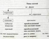
For convenience of study, 5 groups of bones are distinguished by size and shape (Fig. Nos. 22 and 23).
1) Long (tubular) boneshave an elongated middle part of a cylindrical or trihedral shape - the body, or diaphysis; thick ends - epiphyses with articular surfaces; areas where the diaphysis goes into the epiphysis, - metaphysis; the elevations protruding above the surface of the bone are apophyses. Form the skeleton of the limbs.
2) Short (spongy) bones have the shape of an irregular cube or polyhedron, such as the bones of the wrist and the tarsus.
3) Flat bones participate in the formation of body cavities, for example, the bones of the skull roof, pelvic bones, ribs, sternum.
4) Abnormal (mixed) bones, for example, vertebrae: their body in form and structure refers to the spongy bones, the arc and processes - to the flat.
5) Airy bones They have a cavity in the body, lined with mucous membrane and filled with air. These include some of the bones of the skull: the frontal, sphenoid, ethmoid, temporal and maxillary.
The growth of the tubular bone in length is due to the metaphyseal (epiphyseal) cartilage between the epiphysis and diaphysis. Complete replacement of the epiphyseal cartilage with bone tissue and the termination of skeletal growth occurs in men aged 23–25 years, in women - 18–20 years. From this time on, the growth of a person stops. Bone growth in thickness occurs due to the periosteum (periosteum), its cambial layer.
Bone strength is very high. It can be compared with the strength of metal or reinforced concrete. For example, the femur, reinforced with ends on props, can withstand a load of 1200 kg, and the tibia in a vertical position - 1650 kg.
Types of bone compounds
Bone connections(Fig. No. 49) unite the bones of the skeleton into a single whole, keep them next to each other and provide them with greater or lesser mobility, a spring (spring) function, as well as growth of the skeleton and the body of the person as a whole.
There are 3 types of bone compounds (Fig. No. 24):
- continuous (synarthrosis) - ligaments, membranes, sutures (skull bones), hammering (tooth-alveolar joints), cartilaginous synchondrosis(temporary, permanent), bone - synostoses;
- discontinuous (joints, diarthrosis);
- transitional form (half-joint, symphysis, hemiarthrosis).
Continuous bone connections with dense fibrous connective tissue are syndesmosesusing cartilage - synchondrosisusing bone tissue - synostoses. The most advanced forms of connection of bones in the human body are discontinuous connections - joints (diarthrosis). These are mobile connections of bones with each other, in which the function of movement comes to the fore. There are a lot of joints in the human body. There are about 120 of them in one vertebral column. But the plan of the structure of all joints is the same.
In the joint allocate the main and auxiliary elements.
The main elements of the joint include:
1) articular surfaces;
2) articular cartilage;
3) the joint capsule;
4) the articular cavity;
5) synovial fluid.
Ancillary elements of the joint include:
1) ligaments;
2) articular discs;
3) articular menisci;
4) joint lips;
5) synovial bags.
Articular surfaces - These are the areas of contact of articulated bones. They have a different shape: spherical, cup-shaped, elliptical, saddle-shaped, condylar, cylindrical, block-shaped, helical. If the articulated surfaces of the bones correspond in size and shape to each other, then these are congruent (Latin congruens - corresponding, coincident) articular surfaces. If the articular surfaces do not match each other in shape and size, then these are incongruent articular surfaces. Articular cartilage with a thickness of 0.2 to 6 mm covers the articular surfaces and thus smooths bone irregularities and dampens movement. Most articular surfaces are covered with hyaline cartilage. The articular capsule hermetically closes the articular surfaces from the environment. It consists of two layers: the outer - fibrous membrane, very dense and strong, and the inner - synovial membrane that produces fluid - synovium. Articular cavity - it is a narrow gap bounded by articular surfaces and synovial membrane, hermetically isolated from surrounding tissues. Has always negative pressure. Synovial fluid - it is a viscous transparent liquid, resembling egg white, which is located in the cavity of the joint. It is a product of the exchange of the synovial membrane of the capsule and articular cartilage. Plays the role of lubricant and buffer cushion.
Bundles - extra-articular (extra capsule and capsular) and intra-articular - strengthen the joint and the capsule. Articular Discs and Menisci - these are continuous and non-continuous cartilaginous plates, which are located between the (incongruent) articular surfaces that are not fully matched to each other. They smooth the irregularities of articulated surfaces, making them congruent. Articular lip - cartilaginous roller around the articular cavity to increase its size (shoulder, hip joints). Synovial bag - This is the protrusion of the synovial membrane in the thinned areas of the fibrous membrane of the joint capsule (knee joint).
The joints differ from each other in structure, shape of articulated surfaces, range of motion (biomechanics). The joint formed by only two articular surfaces is simple joint; three or more articular surfaces - complex joint. The joint characterized by the presence between the articulated surfaces of the articular disc (meniscus), which divides the joint cavity into two floors, is complex joint. Two anatomically isolated joints acting together constitute combo joint.
Hemiarthrosis (polusustav, symphysis) This is a cartilaginous joint of bones, in which there is a narrow gap in the center of the cartilage. Such a connection is not covered externally by the capsule, and the inner surface of the slit is not lined with synovial membrane. In these compounds, small displacements of bones relative to each other are possible. These include the symphysis of the handle of the sternum, the intervertebral symphysis and the pubic symphysis.
3. Spinal column (Fig. Nos. 25 and 26)
Spinal column, chest and skull belong to axial skeletonThe bones of the upper and lower limbs are called additional skeleton.
Spinal column(Fig. No. 27), or the spine, is located on the back of the body. It performs the following functions:
1) supporting, being a rigid rod that holds the weight of the body;
2) protective, forming a cavity for the spinal cord, as well as the organs of the chest, abdominal and pelvic cavities;
3) locomotor, participating in the movements of the torso and head;
4) spring, or spring, softening the shocks and tremors received by the body when jumping, running, etc.
There are 33-34 vertebra in the vertebral column, of which 24 free are true (cervical, thoracic, lumbar), and the rest are intergrown - false (sacral, coccygeal). There are 7 cervical, 12 thoracic, 5 lumbar, 5 sacral and 4-5 coccygeal vertebrae. True vertebrae have a number of common features. In each of them there is a thickened part - the body facing forward and an arc going backward from the body, limiting the vertebral foramen. When vertebrae connect, these openings form the spinal canal in which the spinal cord is located. Seven processes extend from the arc: one unpaired - spinous facing backwards; the remaining pairs: the transverse processes are directed to the sides of the vertebrae, the upper articular processes go upwards and the lower articular processes are directed downwards. At the junction of the vertebral arch with the body, there are two vertebral notches on each side: upper and lower, which form intervertebral holes when the vertebrae connect. The spinal nerves and blood vessels pass through these openings.
Cervical vertebrae(Fig. No. 28) have the characteristic features that distinguish them from the vertebrae of other departments. The main difference is the presence of a hole in the transverse processes and a split at the end of the spinous processes. The spinous process of the VII cervical vertebra is not split, it is longer than the others and can be easily felt under the skin (protruding vertebra). On the anterior surface of the transverse processes of the VI cervical vertebra there is a well-developed dormant tubercle - a place where the common carotid artery can easily be squeezed to temporarily stop bleeding. I cervical vertebra - atlas It has no body and spinous process, but contains only two arches and lateral masses, on which the articular fossae are located: the upper ones for articulation with the occipital bone, the lower ones for articulation with the II cervical vertebra. II cervical vertebra - axial (epistrophy) - has a toothlike process on the upper surface of the body - a tooth, around which the head rotates (together with the atlas).
Have thoracic vertebrae(Fig. No. 29) the spinous processes are the longest and directed downwards; in the lumbar ones, they are wide in the shape of quadrangular plates and directed straight back. On the body and transverse processes of the thoracic vertebrae there are rib holes for articulation with the heads and tubercles of the ribs.
Sacral bone, or the sacrum, consists of five sacral vertebrae (Fig. Nos. 30 and 31), which by the age of 20 will grow into one monolithic bone, which gives this spinal section the necessary strength.
Coccyx, or the tailbone, consists of 4-5 small underdeveloped vertebrae.
The human spinal column has several bends. The bends facing the bulge forward are called lordosis, the bulge backward is called kyphosis, and the bulge to the right or left is scoliosis. The following physiological curves are distinguished: cervical and lumbar lordosis, thoracic and sacral kyphosis, thoracic (aortic) scoliosis. The latter occurs in 1/3 of cases, located at the level of the III-V thoracic vertebrae in the form of a slight bulge to the right and caused by the passage at this level of the thoracic aorta.
Rib cage
Rib cage(Fig. No. 32), formed by 12 pairs of ribs, sternum and thoracic spinal column. It is the skeleton of the walls of the chest cavity, in which there are important internal organs (heart, lungs, trachea, esophagus, etc.).
SternumThe brisket is a flat bone consisting of three parts: the upper one is the handle, the middle one is the body, and the lower one is the xiphoid process. In newborns, all 3 parts of the sternum are constructed of cartilage, in which the nuclei of ossification are located. In adults, only the arm and body are interconnected by cartilage. By 30-40 years, the ossification of cartilage is completed, and the sternum becomes a monolithic bone. On the upper edge of the handle, there is a jugular notch, and on the sides of it - clavicular cuts. On the outer edges of the body and the handle are seven cutouts for the ribs.
Ribs - These are long flat bones. There are 12 pairs. Each rib has a large posterior bone portion and a smaller anterior cartilage, which grow together. The rib has a head, neck and body. Between the neck and the body, in the upper 10 pairs there is a tubercle of the rib, which has an articular surface for articulation with the transverse process of the vertebra. On the head of the rib there are two articular areas for articulation with the rib pits of two adjacent vertebrae. The ribs distinguish outer and inner surfaces, upper and lower edges. On the inner surface, along the lower edge, a rib groove is visible - a trace of the occurrence of vessels and nerves.
The ribs are divided into three groups. The top 7 pairs of ribs that reach their sternum cartilages are called true. The next 3 pairs, connecting with each other with their cartilage and forming a costal arch, are called false. The last 2 pairs of their ends freely lie in soft tissues, they are called hesitant ribs.
The thorax is generally shaped like a truncated cone. The upper opening of the chest, limited by the body of the I thoracic vertebra, the first pair of ribs and the upper edge of the sternum handle, is free. Through it to the neck of the tops of the lungs, as well as the trachea, esophagus, blood vessels and nerves. The lower opening of the thorax is limited to the body of the XII thoracic vertebra, ribs XI and XII pairs, costal arches and the xiphoid process. This hole is hermetically tightened by the diaphragm. Since the I rib during breathing is very little mobile, therefore, ventilation of the tops of the lungs during breathing is minimal. This creates favorable conditions for the development of inflammatory processes in the apex of the lungs.
Numerous bone connections It is advisable to present in the form of classification. In accordance with this classification, there are two main types of bone compounds - continuous and discontinuous, each of which, in turn, is divided into several groups (Gayvoronsky I. V., Nichiporuk, G. I., 2005).
Types of bone compounds
| Continuous connections (synarthrosis, synarthrosis) | Disconnected joints (diarthrosis, diarthrosis; synovial joints or joints, articulationes synoviales) |
|
I. Fibrous compounds (articulationes librosae): ligaments (ligamenta); membranes (membranae); springs (fonticuli); stitches (suturae); hammering (gomphosis) Ii. Cartilage compounds (articulationes cartilagineae): compounds with hyaline cartilage (temporary); connections using fibrous cartilage (permanent) Iii. Compounds using bone tissue (synostosis) |
Along the axis of rotation and the shape of the articular surfaces: By the number of articular surfaces: simple (art. Simplex); complex (art. composite) Single-step joint function: combined (art. Combinatoria) |
It should be noted that the relief of the bones often reflects the specific type of compound. For continuous joints on bones, tuberosities, ridges, lines, pits and roughness are characteristic, and for discontinuous smooth articular surfaces of various shapes.
Continuous bone connections
There are three groups of continuous joints of bones - fibrous, cartilaginous and bone.
I. Fibrous bone compounds, or compounds with connective tissue, syndesmosis. These include ligaments, membranes, fontanelles, sutures and hammering.
Bundles are joints with connective tissue, having the appearance of bundles of collagen and elastic fibers. By their structure, ligaments with a predominance of collagen fibers are called fibrous, and ligaments containing predominantly elastic fibers are elastic. Unlike fibrous, elastic ligaments are able to shorten and return to their original form after the cessation of stress.
Along the length of the fibers, ligaments can be long (posterior and anterior longitudinal ligaments of the spinal column, supraspastic ligament), connecting several bones over a large distance, and short, connecting adjacent bones (interspinous, intertransverse ligaments and most ligaments of the limbs).
In relation to the joint capsule, intraarticular and extraarticular ligaments are distinguished. The latter are considered as extracapsular and capsular. Bundles as an independent type of bone connection can perform various functions:
- retaining or fixing (sacroiliac ligament, sacrospinous, interosseous, cross-border ligaments, etc.);
- the role of the soft skeleton, as they are the site of the beginning and attachment of muscles (most of the ligaments of the limbs, ligaments of the spinal column, etc.);
- formative when they together with the bones form arches or openings for the passage of vessels and nerves (upper transverse ligament of the scapula, pelvic ligaments, etc.).
Membranes are connections with the help of connective tissue, having the appearance of an interosseous membrane that, unlike the ligaments, fills the gaps between the bones. Connective tissue fibers in the composition of the membranes, mainly collagen, are arranged in a direction that does not impede movement. Their role is in many ways similar to bundles. They also hold the bones relative to each other (intercostal membranes, interosseous membranes of the forearm and lower leg), serve as the starting point of the muscles (the same membranes) and form openings for the passage of blood vessels and nerves (obturator membrane).
Rodnichki are connective tissue formations with a large amount of intermediate substance and rarely located collagen fibers. Springflows create conditions for the displacement of the bones of the skull during childbirth and contribute to the intensive growth of bones after birth. The front fontanel reaches the largest size (30 x 25 mm). It closes in the second year of life. The back fontanel has a size of 10 x 10 mm and disappears completely by the end of the second month after birth. Even smaller sizes have paired wedge-shaped and mastoid springs. They overgrow before birth or in the first two weeks after birth. Springflows are eliminated due to the proliferation of the bones of the skull and the formation between them suture connective tissue.
The seams are thin layers of connective tissue, located between the bones of the skull, with a large number of collagen fibers. The seams are serrated in shape, scaly and flat, they serve as a zone of growth of the bones of the skull and have a cushioning effect during movements, protecting the brain, organs of sight, hearing and balance from damage.
Insertion - the connection of the teeth with the cells of the alveolar processes of the jaws with the help of dense connective tissue, which has a special name - periodontal. Although it is a very strong compound, it also has pronounced damping properties under the load on the tooth. The thickness of the periodontium is 0.14–0.28 mm. It consists of collagen and elastic fibers, oriented all along perpendicular from the walls of the alveoli to the root of the tooth. Between the fibers lies loose connective tissue containing a large number of vessels and nerve fibers. When the jaws are strongly compressed due to the pressure of the antagonist tooth, the periodontium is strongly compressed, and the tooth is immersed in the cell to 0.2 mm.
With age, the number of elastic fibers decreases, and under load the periodontium is damaged, its blood supply and innervation are disturbed, the teeth become loose and fall out.
Ii. Bone cartilage - synchondrosis. These compounds are represented by hyaline or fibrous cartilage. Comparing these cartilages with each other, it can be noted that hyaline cartilage is more elastic, but less durable. Metaphysis and epiphysis of tubular bones and separate parts of the pelvic bone are connected with the help of hyaline cartilage. Fibrous cartilage is mainly composed of collagen fibers, therefore, is more durable and less elastic. This cartilage connects the vertebral body. The strength of cartilaginous joints is also increased due to the fact that the periosteum from one bone passes to another, without interrupting. In the area of cartilage, it turns into a supragrass, which in turn firmly fuses with cartilage and is supported by ligaments.
For the duration of existence of synchondrosis can be permanent and temporary, i.e., existing up to a certain age, and then replaced by bone tissue. Under normal physiological conditions, the metaepiphyseal cartilage, the cartilage between the individual parts of the flat bones, the cartilage between the main part of the occipital and the body of the sphenoid bones are temporary. These compounds are mainly represented by hyaline cartilage. Constant are cartilage forming intervertebral discs; cartilage, located between the bones of the base of the skull (wedge-stony and wedge-occipital), and cartilage between the I rib and sternum. These compounds are represented mainly by fibrous cartilage.
The main purpose of synchondrosis is to mitigate jolts and stresses under heavy loads on the bone (depreciation) and to ensure a strong connection of the bones. Cartilaginous compounds at the same time have great mobility. The range of movements depends on the thickness of the cartilaginous layer: the larger it is, the greater the volume of movement. As an example, you can take a variety of movements in the spinal column: bending forward, backward, sideways, twisting, springing movements, which are especially developed in gymnasts, acrobats and swimmers.
Iii. Bone Tissue Compounds - synostoses. These are the most durable compounds from the group of continuous, but completely lost elasticity and depreciation properties. Under normal conditions, temporary synchondrosis undergo synostosis. In some diseases (Bechterew's disease, osteochondrosis, etc.), ossification can occur not only in all synchondrosis, but also in all syndesmosis.
Disconnected bone connections
Disconnected joints are joints or synovial joints. A joint is a discontinuous abdominal joint formed by articulating articular surfaces, covered with cartilage, enclosed in an articular bag (capsule), inside which contains synovial fluid.
The joint must necessarily include three main elements: the articular surface covered with cartilage; articular capsule; joint cavity.
1. Articular surfaces - These are bone areas covered with articular cartilage. In the long tubular bones they are located on the epiphyses, in the short ones - on the heads and bases, in the flat ones - on the processes and the body. The forms of the articular surfaces are strictly deterministic: more often there is a head on one bone, a fossa on the other, less often they are flat. The articular surfaces on the articulated bones must be consistent in shape, that is, be congruent. Often the articular surfaces are lined with hyaline (vitreous) cartilage. The fibrous cartilage covers, for example, the articular surfaces of the temporomandibular joint. The thickness of the cartilage on the articular surfaces is 0.2-0.5 cm, and in the articular fossa it is thicker along the edge, and at the articular head in the center.
In the deep layers, the cartilage is calcified, firmly connected with the bone. This layer is called omelioration, or impregnated with calcium carbonate. Chondrocytes (cartilaginous cells) in this layer are surrounded by connective tissue fibers located perpendicular to the surface, i.e., in rows or columns. They are adapted to resist the pressure forces on the articular surface. In the surface layers of the connective tissue fibers in the form of arcs, starting and ending in the deep layers of cartilage. These fibers are oriented parallel to the surface of the cartilage. In addition, in this layer there is a large amount of intermediate substance, so the surface of the cartilage is smooth, as if polished. The surface layer of cartilage is adapted to the resistance to friction forces (tangential forces). With age, the cartilage undergoes making of wood, its thickness decreases, it becomes less smooth.
The role of articular cartilage is reduced to the fact that it smoothes the irregularities and roughness of the articular surface of the bone, giving it greater congruence. Because of its elasticity, it softens shocks and tremors, so the articular cartilage is thicker in the joints that carry a large load.
2. Articulated bag - it is a hermetic capsule that surrounds the articular cavity, which grows along the edge of the articular surfaces or at a small distance from them. It consists of the outer (fibrous) membrane and the inner (synovial). The fibrous membrane, in turn, consists of two layers of dense connective tissue (outer longitudinal and inner circular), in which the blood vessels are located. It is reinforced by extra-articular ligaments, which form local thickenings and are located in places of greatest load. Bundles are usually closely connected with the capsule, and they can only be separated artificially. Rarely are ligaments detached from the joint capsule, for example, the lateral large tibia and peroneal. In sedentary joints, the fibrous membrane is thickened. In the movable joints, it is thin, loosely stretched, and in some places is so much thinned that the synovial membrane protrudes. This forms synovial inversions (synovial bags), usually located under the tendons.
The synovial membrane is facing the joint cavity, is abundantly supplied with blood, is lined from the inside by synoviocytes capable of secreting synovial fluid. The synovial membrane covers the inside of the entire joint cavity, goes to the bones and intra-articular ligaments. Only surfaces represented by cartilage remain free of it. The synovial membrane is smooth, shiny, can form numerous processes - villi. Sometimes these villi come off and how foreign bodies fall on the inter-articular surfaces, causing short-term pain and impeding movement. This condition is called the "articular mouse." The synovial membrane may lie directly on the fibrous membrane or be separated from it by the subynovial layer or fat layer, therefore, fibrous, areolar and fatty synovial membranes are distinguished.
The synovial fluid is a transudate in terms of the composition and nature of the formation — the effusion of blood plasma and lymph from the capillaries adjacent to the synovial membrane. In the cavity of the joint, this fluid mixes with the debris of rejected synoviocyte cells and abrading cartilage. In addition, the composition of the synovial fluid includes mucin, mucopolysaccharides and hyaluronic acid, which give it viscosity. The amount of fluid depends on the size of the joint and ranges from 5 mm3 to 5 cm3. Synovial fluid performs the following functions:
- lubricates articular surfaces (reduces friction during movement, increases glide);
- couples articular surfaces, keeps them relative to each other;
- softens the load;
- nourishes articular cartilage;
- involved in metabolism.
3. Joint cavity - it is a hermetically closed space bounded by articular surfaces and a capsule, filled with synovial fluid. It is possible to isolate the joint cavity on an intact joint only conditionally, since there are no voids between the articular surfaces and the capsule, it is filled with synovial fluid. The shape and volume of the cavity depends on the shape of the articular surfaces and the structure of the capsule. In sedentary joints, it is small, in high-mobility - large and may have eversion extending between bones, muscles and tendons. In the cavity of the joint pressure is negative. When the capsule is damaged, air penetrates into the cavity, and the articular surfaces diverge.
In addition to the main elements, there may be auxiliary joints in the joints, which ensure optimal joint function. These are intra-articular ligaments and cartilage, articular lips, synovial folds, sesamoid bones and synovial bags.
- Intra-articular ligaments- these are fibrous ligaments, covered with a synovial membrane, which bind the articular surfaces in the knee joint, in the joint of the head of the rib and the hip joint. They hold the articular surfaces relative to each other. This function is particularly clearly seen in the example of the cruciate ligament of the knee joint. When they break, a “drawer” symptom is observed, when, when bent at the knee joint, the lower leg shifts towards the femur anteriorly and posteriorly by 2-3 cm. The ligament of the femoral head serves as a conduit for the vessels feeding the articular head.
- Intra-articular cartilage - This is fibrous cartilage, located between the articular surfaces in the form of plates. The plate completely dividing the joint into two "floors" is called the articular disc (discus articularis). In this case, two separated cavities are formed, such as, for example, in the temporomandibular joint. If the joint cavity is separated only partially by the plates of the cartilage, that is, the plates have the shape of a semi-moon and are spliced with the capsule, these are menisci, which are presented in the knee joint. Intra-articular cartilages ensure the congruence of the articular surfaces, thereby increasing the range of movements and their diversity, help to soften the shocks, reduce the pressure on the underlying articular surfaces.
- Articular lip - it is a fibrous cartilage of a ring-shaped form, supplementing the articular fossa along the edge; at the same time, one lip is spliced with the joint capsule, and the other one passes into the articular surface. The articular lip is found in two joints: shoulder and hip (labrum glenoidale, labrum acetabulare). It increases the area of the articular surface, makes it deeper, thereby limiting the range of motion.
- Synovial folds (plicae synoviales) - it is a vessel-rich connective tissue formation, covered with synovial membrane. If fatty tissue accumulates inside them, fat folds are formed. The folds fill the free space of the joint cavity, which has a large size. By helping to reduce the joint cavity, the folds indirectly increase the adhesion of the articulated surfaces and thereby increase the range of motion.
- Sesamoid bones (ossa sesamoidea)- These are intercalated bones, closely connected with the joint capsule and the surrounding tendons of the muscles. One of their surfaces is covered with hyaline cartilage and facing the joint cavity. Inserted bones help to reduce the joint cavity and indirectly increase the range of motion in it. They are also blocks for the tendons of the muscles acting on the joint. The largest sesamoid bone is the patella. Small sesamoid bones are often found in the joints of the hand, foot (in the interphalangeal, carpal-metacarpal joint of the 1st finger, etc.).
- Synovial bags (bursae synoviales) - These are small cavities lined by the synovial membrane, often communicating with the joint cavity. Their size is from 0.5 to 5 cm3. A large number of them found in the joints of the limbs. Inside them, synovial fluid accumulates, which lubricates the adjacent tendons.
Movement in the joints can be carried out only around three axes of rotation:
- frontal (axis corresponding to the frontal plane dividing the body into front and rear surfaces);
- sagittal (axis corresponding to the sagittal plane separating the body into right and left halves);
- vertical, or its own axis.
For the upper limb, the vertical axis passes through the center of the humeral head, the condyle of the humerus, and the head of the radius and ulna. For the lower limb, in a straight line connecting the anterior superior iliac spine, the inner edge of the patella and the thumb.
The articular surface of one of the articulated bones, having the shape of a head, can be represented as a ball, ellipse, saddle, cylinder, or block. Each of these surfaces corresponds to the articular fossa. It should be noted that the articular surface can be formed by several bones, giving it a certain shape as a whole (for example, the articular surface formed by the bones of the proximal wrist).
1 - ellipsoidal; 2 - saddle; 3 - spherical; 4 - block; 5 - flat
Movement in the joints around the axis of rotation is determined by the geometric shape of the articular surface. For example, a cylinder and a block rotate only around one axis; ellipse, oval, saddle - around two axes; ball or flat surface - around three.
The number and possible types of movements around existing axes of rotation are presented in the tables. Thus, around the frontal axis, two types of movements are noted (flexion and extension); around the sagittal axis there are also two types of movements (adduction and abduction); when moving from one axis to another, another movement occurs (circular or conical); around the vertical axis - one movement (rotation), but it may have some subspecies: rotation in or out (pronation or supination).
The axis of rotation, the number and types of possible movements
The maximum number of possible types of movements in the joints, depending on the number of axes of rotation and the shape of the articular surface
| Osnost joint | The shape of the articular surface | Realized axis of rotation | Number of movements | Types of movements |
| Uniaxial | Blocky | Frontal | 2 | Flexion, extension |
| Rotational (cylindrical) | Vertical | 1 | Rotation | |
| Biaxial | Ellipsoid, saddle | Sagittal and frontal | 5 | Flexion, extension, adduction, abduction, circular motion |
| Condylar | Frontal and vertical | 3 | Flexion, extension, rotation | |
| Multiaxial | Spherical, flat | Frontal, sagittal and vertical | 6 | Flexion, extension, adduction, abduction, circular motion, rotation |
Thus, there are only 6 types of movements. Additional movements are also possible, such as sliding, springing (removal and drawing together of articular surfaces under compression and tension) and twisting. These movements are not related to individual joints, but to a group of combined, for example, intervertebral.
Based on the classification of the joints, it is necessary to characterize each individual group.
I. Classification of joints according to the axes of rotation and the shape of the articular surfaces:
Uniaxial joints- these are joints in which movements are made only around any one axis. Practically, such an axis is either frontal or vertical. If the axis is frontal, then in these joints movements are made in the form of flexion and extension. If the axis is vertical, then only one movement is possible - rotation. Representatives of uniaxial joints in the form of articular surfaces are: cylindrical (articulatio trochoidea) (rotational) and blovidny (ginglymus). Cylindrical joints move around a vertical axis, i.e. they rotate. An example of such joints are: median atlantoaxial joint, proximal and distal radioulnar joints.
The block-like joint is similar to a cylindrical one, only located not vertically, but horizontally and has a comb on the articular head, and a notch on the articular fossa. Due to the scallop and the notch, it is not possible to shift the articular surfaces to the sides. The capsule in such joints is free in front and behind and is always strengthened by lateral ligaments that do not interfere with movements. The block-like joints always work around the frontal axis. An example is interphalangeal joints.
A type of block-like joint is the cochlear (articulatio cochlearis), or helical, joint, in which the notch and scallop are chamfered, have a helical stroke. An example of the cochlear joint is the shoulder joint, which also works around the frontal axis. Thus, uniaxial joints have one or two types of movement.
Biaxial joints - joints working around two of the three available axes of rotation. So, if movements are made around the frontal and sagittal axes, then such joints implement 5 types of movements: flexion, extension, adduction, abduction and circular movement. The shape of the articular surfaces of these joints are ellipsoidal or saddle-shaped (articulatio ellipsoidea, articulatio sellaris). Examples of ellipsoidal joints: atlantococyclic and wrist; saddle: carpometacarpal joint of the 1st finger.
If the movements are carried out around the frontal and vertical axes, then it is possible to realize only three types of movements - flexion, extension and rotation. In shape, these are condylar joints (articulatio bicondyllaris), such as the knee and temporomandibular joints.
Condylar joints are a transitional form between uniaxial and biaxial joints. The main axis of rotation in them is the front. In contrast to uniaxial joints, the difference in areas of the articular surfaces is greater in them, and as a result, the range of movements increases.
Multiaxis joints - these are joints, movements in which are carried out around all three axes of rotation. They make the maximum possible number of movements - 6 types. In shape, these are spherical joints (articulatio spheroidea), such as the shoulder. A type of spherical joint is a cup-shaped (articulatio cotylica), or nut-shaped (articulatio enarthrosis), for example, hip. It is characterized by a deep articular fossa, a strong capsule, strengthened by ligaments, the volume of movements in it is less. If the surface of the ball has a very large radius of curvature, then it approaches a flat surface. A joint with such a surface is called flat (articulatio plana). For flat joints, a small difference in the areas of the articular surfaces, strong ligaments, and movements in them are sharply limited or absent altogether (for example, in the sacroiliac joint). In this regard, these joints are called sedentary (amphiarthrosis).
Ii. Classification of joints by the number of articular surfaces.
Simple joint (articulatio simplex) - This is a joint that has only two articular surfaces, each of which can be formed by one or several bones. For example, the articular surfaces of the interphalangeal joints are formed only by two bones, and one of the articular surfaces in the wrist joint is formed by the three bones of the proximal wrist.
Difficult joint (articulatio composita)- it is a joint, in one capsule of which there are several articular surfaces, therefore, several simple joints, capable of functioning both together and separately. An example of a complex joint is the elbow joint, which has 6 separate articular surfaces, which form 3 simple joints: brachioradial, brachial and octopus, proximal radioulnacular. Some authors also include the knee joint in complex joints. Given the articular surfaces on the meniscus and the patella, they secrete such simple joints as the femoral-meniscus, meniscal-tibial and femoral-patellar. We consider the knee joint to be simple, since the menisci and the patella are auxiliary elements.
Iii. Classification of joints by simultaneous joint function.
Combined joints (articulatio combinatoria) - these are joints that are anatomically disconnected, that is, they are located in different joint capsules, but function only together. For example, temporomandibular joint, proximal and distal radioulnar joints. It should be emphasized that in true combination joints one cannot make movement only in one of them, for example, only in one temporomandibular joint. With a combination of joints with various forms of articular surfaces, movements are realized along a joint having a smaller number of axes of rotation.
Factors determining the amount of movement in the joints.
- The main factor is the difference in the area of articulated articular surfaces. Of all the joints, the largest difference is the area of the articular surfaces in the shoulder joint (the area of the head of the humerus is 6 times the area of the articular cavity on the scapula), therefore in the shoulder joint there is the largest amount of movement. In the sacroiliac joint, the articular surfaces are equal in area, so there is practically no movement in it.
- The presence of auxiliary elements. For example, menisci and discs, increasing the congruence of the articular surfaces, increase the range of motion. Articular lips, increasing the area of the articular surface, contribute to the restriction of movements. Intra-articular ligaments restrict movement only in a certain direction (the cruciate ligaments of the knee joint do not prevent flexion, but oppose excessive extension).
- The combination of joints. For combined joints, movements are determined by a joint having a smaller number of axes of rotation. Although many joints, based on the shape of the articular surfaces, are capable of performing a larger range of movements, it is limited in them because of the combination. For example, in the form of articular surfaces, the lateral atlanto-axial joints are flat, but as a result of combination with the medial atlanto-axial joint, they work as rotational ones. The same applies to the joints of the ribs, hand, foot, etc.
- The condition of the joint capsule. With a thin, elastic capsule movements are made in greater volume. Even the uneven thickness of the capsule in the same joint affects its work. For example, in the temporomandibular joint, the capsule is thinner in front than in the back and side, therefore the greatest mobility in it is anteriorly.
- Strengthening the joint capsule ligaments. Ligaments have a braking and guiding effect, since collagen fibers have not only great strength, but also low tensile properties. In the hip joint, the ilio-femoral ligament prevents the extension and rotation of the limb medially, the pubic-femoral ligament prevents retraction and rotation to the outside. The most powerful ligaments are located in the sacroiliac joint, so there are practically no movements in it.
- The muscles surrounding the joint. Having a constant tone, they fasten, bring together and fix articulated bones. The strength of muscle traction is up to 10 kg per 1 cm2 of muscle diameter. If you remove the muscles, leave the ligaments and the capsule, the range of motion increases dramatically. In addition to the direct inhibitory effect on movements in the joints, the muscles also have an indirect effect through the ligaments from which they start. Muscles in their reduction make the ligaments unyielding, elastic.
- Synovial fluid. It has a cohesive effect and lubricates the articular surfaces. With arthrosis-arthritis, when the release of synovial fluid is disturbed, pain, crunching appear in the joints, the range of movements decreases.
- Screw deflection It is only in the shoulder joint and has an inhibitory effect on movement.
- Atmosphere pressure. It contributes to the contact of the articular surfaces with a force of 1 kg per 1 cm2, has a uniform tightening effect, therefore, moderately restricts movement.
- The state of the skin and subcutaneous fat. In obese people, the amount of movement is always less due to the abundant subcutaneous fatty tissue. In slender, toned, in athletes movements are made in greater volume. In case of skin diseases, when elasticity is lost, movements are sharply reduced, and often after severe burns, contractures are formed, which also significantly impede movements.
To determine the range of motion in the joints there are several techniques. Traumatologists determine it with a protractor. For each joint, defined their original position. The starting position for the shoulder joint is the position of the arm that hangs freely along the torso. For the elbow joint - full extension (180 °). Pronation and supination are determined when the elbow joint is bent at a right angle and when the hand is installed in the sagittal plane.
In anatomical studies, the angle of mobility can be calculated from the difference in the arcs of rotation on each of the articulated articular surfaces. The magnitude of the angle of mobility depends on a number of factors: gender, age, degree of training, individual characteristics.
Joint diseases
IN AND. Mazurov
§21.Skeleton of man. Structure, chemical composition and connection of bones
The value of the musculoskeletal system. Bones and muscles belong to the musculoskeletal system (Scheme 2). Connecting to each other with the help of joints and cartilage, the bones form the skeleton of a person. It serves as a support for the body. Muscles are attached to the bones of the skeleton. This is the active part of the oiorno-motor apparatus. Movements are carried out by reducing them. As a result, both the individual bones and the whole body move.
In addition to performing the support function, the bones of the skeleton protect the internal organs from mechanical damage. For example, the brain is protected by the skull bones firmly connected to each other. Chest bones protect the heart and lungs.
Scheme 2
Oporio-implantation system (SLM)
L_______________________________________________
skeleton bones_____________________ skeletal muscle
1. HematopoieticFunctions: 1. Providing movement
2. Support 2. Protection of internal organs
3. Protective (abdominals)
Connecting -Type of fabric - Muscle striated
In the red bone marrow, filling the cancellous bone, blood cells are formed. Since bones contain many minerals (phosphorus, calcium), they are involved in the metabolism.
The human skeleton (Scheme 3) includes the skeleton of the head, or the skull, the skeleton of the body, the skeleton of the upper limbs and the skeleton of the lower extremities (Fig. 50). In the composition of the skeleton of an adult about 220 bones. The bones differ from each other in shape and structure (Scheme I). According to the structure, there are three types of bones: tubular, flat and mixed (vertebrae). Amongtubular bones there are long (humeral, femoral, forearm and lower leg bones) and short (phalanxes of fingers). The cavity of the tubular bones in children is filled with red bone marrow, which is replaced by yellow throughout life.
(adipose tissue).
Haveflat bones length and width is different. These include the scapula, skull bones, sternum, pelvic bones. Flat bones

One should not forget that the long flat bones, such as the ribs, and the short tubular ones are the palns (phalanx) bones.
Participate in the formation of limb girdles and perform the function of protection (bones of the skull, sternum, ribs).
Bone structure Bones are formedbone tissue which is a type of connective tissue (Fig. 51). It is composed of cells n dense intercellular substance. Most bones consist of an outercompact (dense) and internalspongy substance. It is located on the bodies of the flat and in the heads of the tubular bones. Spongy substance consists ofcrossbar located in an arcuate manner, according to the directions in which the bone is under mechanical stress.

Fig. 50. Human skeleton: / skull bones;2 collarbone;
1 - scapula;
2 sternum; 5 - ribs;
6spine:
7pelvic bone;8 - brachial bone;
9 ulna n radius;10 - wrist and hand bones;II - femur:12 - patella (patella):13 - tibia;14 - small tibia:15 - bone moans
Outside, the bone is covered with a periosteum (excluding articular surfaces), which is penetrated by blood vessels that feed the bone. There are many sensory nerve endings in the periosteum. Due to cell division of the periosteum, the bone grows thick and recovers when injured. The ability of bone cells to recover (regenerate) allows the bones to grow together with fractures. The growth of the bones in length contribute to cartilage

Fig. 51. Bone structure:
I bone cells(increased h.); 2 Bone marrow(uvslich.): 3 sponge substance:4 - yellow bone marrow.5- 6 blood vessels:7 - tight in:8 - periosteum
fabrics (a type of connective tissue). Ossification of the body occurs in 20-25 years. Therefore, a person grows up to 25 years.
The composition of the bones. Bones are made up of organic and inorganic substances. Waters in them are 50%, proteins (ossein) - 12.5%, fats 15.7%, mineral substances (calcium, etc.) - 21.8%. Organic matter ossein gives bones strength and flexibility. In the body of children, organic matter is more, so the bones have elastic and elastic. In ballet and circus schools, as well as sports sections, children are admitted from 1-7 years old. With age, the composition of bones decreases the amount of organic matter. The bones lose their plasticity and become more brittle.
The connection of the bones. The bones of the skeleton are interconnected in various ways. There are 3 types of connections for the performed guns: fixed, semi-mobile and mobile.
Fixed connection formed by accretion of bones. This is a bone seam. In this case, the protrusions of one bone grow into the recesses of the other. So bones of a skull are connected (see fig. 63).
Semi movable joint - This is the connection of bones with cartilage. For example, connecting the vertebrae to each other provides the flexibility of the spine (see Fig. 58).
Mobile connection (fig. 52) is the joining of bones with the help of joints. The joint connects the bones of those parts of the skeleton where increased mobility is required - limbs (fig. 53,54), the connection of the skull with the spine. Joints must include the following elements: articular cavity of one bone: head of another bone; articular bag: intraarticular ligaments: articular fluid.
Fig. 52. Mobile connection * of bones in the hip sustiva: |

The fluid acts as a lubricant. It also reduces friction and provides sliding of the articular surfaces of the bones during movement. The amount of joint fluid filling the narrow gap between the joint surfaces is very small. Ligaments (fig. 55, 56) increase the strength of the bonding of parts of the skeleton, limit the amplitude of movements, etc. Movement in the joints is performed by the muscles.
1 4 wrist ligaments:5-6 - palmar ligaments;7 metacarpals;8 metacarpal ligaments;9 metacarpophalangeal joint of the fifth finger
1 anterior interfacial ligament:
2 heel-fibular ligament:
3 - deltoid ligament:4 tarsal ligament;5 metatarsal ligaments;6 joints and ligaments
4
- head humeral to * stn; 5 - tendon of the head of the biceps of the shoulder
The joints are distinguished by their number (simple and complex), the shape of the articular surfaces of the bones (for example, the flat inter-carpal and spherical humeral) (Fig. 57) and the possible range of motion.
Bone tissue, cartilage tissue, compact (dense) substance. spongy substance, periosteum, ossein: immobile
joint suture), semi mobile and mobile joints of bones: joints. articular cavity, articular bag, articular fluid
bone: ligaments.
1. What tissue is the bone made of? Until years
skeleton?
2.What are the properties of iridium organic matter?
3.What types of bone connections are there? Characterize them.
1.What is a joint? Tell us about its structure and functions.
2.Explain how the bone grows thick.
Connection type
Where is the meeting *1 is
Determination of the chemical composition of the bone.
Objective: determine the presence of organic and inorganic substances in the composition of bones.
Equipment: fish ribs, tubular bones of a chicken, small bones of a rabbit; matches; cold ox; hydrochloric or sulfuric acid; a cup with a wide neck.
Working process. The teacher in advance (2-3 days) puts ribs and tubular bones in a 10% solution of hydrochloric or sulfuric acid. During the session, the bones are removed with tweezers and washed in cold water. Try to bend them and make knots of them. Dry bones try to burn.Findings. Write in the notebook what changes occur to the bones that were in the acid. How did the properties of the bone change after burning? Note that when burning organic matter is charred. When lowering in acid from the bones, minerals are removed. What properties give bones organic and inorganic substances?
The human skeleton consists of 206 bones that are interconnected. The bone structure of the trunk, upper and lower extremities is very plastic, which ensures its flexibility and mobility. Such functions work due to different joints of the bones, which perform the function of fixing some parts of the skeleton, as well as the mobility of others.
General information about the human bone apparatus
All bones and their compounds present in the human body can be divided into the following types:
Tubular. The task of the tubular bones is to support the body, providing it with movement. The shape of the tubular bone - a tube that has a bone marrow channel inside. The tubular bone can be long or short.
Spongy. Spongy bone can also be long or short, as well as sesamoid. The functions of the long spongy bones are the support, the protection, while the short bones are only the support. Sesamoid bone is localized in the depth of the tendons, helps to strengthen them.
Flat. The task of flat bones is the formation of a wall between the internal organs. On the one hand, the shape of such a bone is curved, on the other - convex.
Mixed. The presence of mixed bones is observed in the area of the skull.
The surface of each individual bone is not the same and is different in each case. So, the nerves and vessels that are adjacent to the bone can leave grooves, cuts, holes, roughness, channels. In those places where the bones do not attach muscles and ligaments, the surface is ideally smooth. When strong muscles are attached to the surface, it is more bumpy. As an example, we can mention those professions that are associated with heavy physical exertion. People who engage in similar activities have more uneven bone surfaces.
In addition to the connecting surface, the bone has a periosteum - a thin shell, characterized by a multitude of nerves and blood vessels. The periosteum performs the function of feeding the bone.
Bone structure
Human bones are endowed with a complex structure and complex chemical composition.
An adult's bone contains up to 50% water, 28.15% organic and 21.85% inorganic substances.
Inorganic matter contains calcium, phosphorus, magnesium and other elements.
If organic matter is predominant in bone, it becomes significantly resilient and resilient. If inorganic compounds prevail, which is observed in old age in the presence of certain pathologies, there is an increased fragility and fragility of the bone. 
What types of compounds exist?
There are 3 main types of connection between human bones:
- Continuous.
- Semi-continuous.
- Discontinuous.
Continuous connections
Such a connection between the bones provides connective tissue. There are such types of fabric for connection:
- fibrous connective tissue;
- cartilaginous connective tissue;
- bone.
Fibrous types of bone joints between each other include:
Sutures - connective tissue (layer), which is localized between the bones of the skull. The seams are divided into 3 types: flat (zone of location - the facial section of the skull), dentate (brain section), scaly (temporal, parietal bones). The layer absorbs shocks, tremors in the case of walking, jumping. In the old age, most of them overgrow, which can be a source of cranial deformities. If the process of overgrowth of seams does not occur synchronously, this is the way to the appearance of skull asymmetry. 
Tooth-alveolar connective tissue performs the function of connecting the root of the tooth with the walls of the alveoli together.
Syndesmous connective tissue is ligaments and interosseous membranes. The former perform the function of connecting adjacent bones together, directing, limiting their movement, and also strengthening joints. The function of intercostal membranes is the connection of tubular bones to each other.
The cartilaginous connective tissue is very strong as well as elastic. This type of fabric, in turn, is divided into permanent and temporary. Permanent cartilage tissue is present in a person throughout life, and temporary cartilage is replaced with bone tissue over age.
Semi-continuous connections
Semi-continuous connective tissue or, in other words, half-joints - is a compound with the help of cartilage. The cartilage, in turn, contains a slit-like cavity in which there is a fluid that increases the movement of the joint.
Semi-continuous connective tissue - the pubic symphysis, in which two pelvic bones interconnect. The slight divergence of these two bones plays a big role in childbirth in women.
Disconnected connections
Disconnected bone joints are joints and synovial joints.
The joint is the connective tissue that provides mobility, as well as a variety of movements. The scheme of the components is as follows:
Joint surface
The surface is a portion of bone that is covered with cartilage. Basically, the surface of the joint, which are located next to each other, are mutually located. In other words, if the first surface is convex, then the second will be concave. But there are such joints, on the surfaces of which there is a discrepancy between their shape and size.
Cartilage
Cartilage is strongly associated with bone. Its functions are reduced to ensure the smoothing of irregularities, surface roughness. Due to its elasticity, cartilage acts as a means of softening shocks, tremors.
Capsule
The capsule is an envelope surrounding the cavity. It is possible to note its strong accretion with the periosteum, which forms a closed cavity. The outer layer of the capsule is the fibrous membrane, and the inner layer is the synovial membrane.
Cavity
The cavity contains synovial fluid in its space. 
Intra-articular fluid
The functions of the intra-articular fluid are reduced to a reduction in friction of the surfaces of the joints in the case of movement, an increase in sliding, adhesion of surfaces, a softening of the load, and nutrition of cartilage.
In addition to the basic elements, there are also auxiliary ones. These include:
Ligaments - some brakes that limit the mobility of the joint. There are extra-articular and intra-articular ligaments.
Disk (dividing the joint into two halves), menisci (reducing the pressure on the joint).
The lip performs the function of increasing the surface area of the joint, making it deeper.
Synovial folds fill the free space of the cavity. With the accumulation of fatty tissue, the formation of fat folds.
Sesamoid bones are closely related to the capsule. Their function is to reduce the joint cavity, increase the range of motion of the joint.
A synovial bag is a cavity that has synovial fluid in its space. This fluid is a lubricant for adjacent tendons. 
Connective tissue pathology
There are such types of diseases in humans:
Hereditary type
The occurrence of the disease in humans is associated with a change in a particular gene. This reason for the development of pathologies is quite rare, but still occurs. These include the Ehlers-Danlos syndrome. Symptoms of pathology consist in the curvature of the spine, weakness of blood vessels, bleeding gums, impaired functioning of the lungs, heart, and digestive system. Other diseases can be called:, teeth fragility.
Autoimmune type
The cause of the occurrence of an autoimmune human disease is considered some kind of antibody fight with the body’s own tissues. These diseases include: polymyositis, dermatomyositis. The common symptoms of such pathologies are muscular weakness, fatigue, shortness of breath, and weight loss. Dermatomyositis is accompanied by damage to the skin around the eyes, as well as in the area of the palms.
One of these diseases in humans is rheumatoid arthritis. In this case, a lesion of the membrane that surrounds the joint occurs. Symptoms of the disease are: pain, reduced range of motion, swelling, inflammation process. Common symptoms for all: fatigue, anemia, fever, loss of appetite. 
Systemic lupus is considered an autoimmune disease, during which immunity suppresses its own connective tissue cells. Cases of transmission of the disease from one person to another are not fixed. The development of this disease occurs at the genetic level. But there are some suggestions why lupus erythematosus can occur:
- under the influence of ultraviolet radiation;
- smoking;
- infections present;
- exposure to chemicals.
Acceptance of certain drugs (antipsychotic, biological, hormonal, anti-inflammatory, antifungal).
Diagnosing systemic lupus is quite difficult, since its manifestation may be similar to a different pathology. The most distinctive feature is erythema on the face. The following symptoms are also present:
- fast fatiguability;
- elevated temperature;
- pain syndrome, joint swelling;
- chest pains;
- memory impairment.
 The doctor selects the necessary treatment, given the severity of symptoms. The use of nonsteroidal drugs, antimalarial drugs, corticosteroids, immunosuppressants is recommended.
There are cases when a person has several pathologies of connective tissue at the same time. It may be lupus, polymyositis, dermatomyositis, rheumatoid arthritis. These diseases are the cause of the diagnosis, which is called "mixed connective tissue disease." Some people do not complain about the inconvenience and discomfort, but the complications can be quite dangerous: infection, renal failure, heart attack, stroke.
Treatment of a mixed disease, like any other, should be prescribed only by a doctor who takes into account the course of the pathology, the severity of the symptoms, their severity.
Classification of compounds. There are two main types of bone connections: continuous and discontinuous, or joints.Continuous compounds are present in all lower vertebrates and in embryonic developmental stages in higher ones. When the bone marrow forms are formed in the latter, their source material (connective tissue, cartilage) is preserved between them. With the help of this material the bones are bonded, i.e. formed a continuous connection. Disconnected connections develop at later stages of ontogenesis in terrestrial vertebrates and are more perfect, since they provide a more differentiated mobility of parts of the skeleton. They develop as a result of the occurrence of a gap in the original material that has been preserved between the bones. In the latter case, cartilage residues cover the articulating surfaces of the bones. There is a third, intermediate type of compounds - half-articulate
Continuous connections. Continuous connection - synarthrosis, or fusion, occurs when the bones are connected to each other by a connecting tissue. The movements are extremely limited or absent. By the nature of the connective tissue, connective tissue adhesions are distinguished, or syndesmoses , cartilaginous adhesions, or synchondrosis , and adhesions with the help of bone tissue - synostoses.
Syndesmoses There are three kinds: 1) interosseous membranes, for example between the bones of the forearm or
lower leg; 2) bundles, connecting bones (but not related to the joints), for example, ligaments between the processes of the vertebrae or their arches; 3) stitches between the bones of the skull.
Types of bone connections (scheme):
BUT - syndesmosis; B - synchondrosis; AT - joint; 1 - the periosteum; 2 - bone; 3 - fibrous connective tissue; 4 - cartilage; 5 - synovial and 6 - fibrous layer of the articular bag; 7 - articular cartilage; 8 - joint cavity
Interosseous membranes and ligaments allow some displacement of the bones. In the seams the interlayer of connective tissue between the bones is very small and movement is impossible.
Synchondrosis is, for example, the connection of the I rib to the sternum by means of costal cartilage, the elasticity of which allows for some mobility of these bones.
Synostoses develop from syndesmoses and synchondrosis with age, when the connective tissue or cartilage between the ends of some bones is replaced by bone tissue. An example is the fusion of the sacral vertebrae and overgrown sutures of the skull. There are naturally no movements here.
Disconnected connections. Disconnected - diarthrosis, articulation, or joint , characterized by a small space (gap) between the ends of the connecting bones. There are joints simple, formed by only two bones (for example, the shoulder joint), complex ones - when more bones (for example, the elbow joint) are included in the joint, and combined, allowing movement only simultaneous with movement in other anatomically isolated joints (for example, proximal and distal radioulnar joints). The composition of the joint includes: articular surfaces, articular bag, or capsule, and articular cavity.
Articular surfaces connecting bones more or less correspond to each other (congruent). On one bone that forms the joint, the articular surface is usually convex and is called heads. The corresponding concavity of the head develops on the other bone - hollow or fossa. Both the head and the fossa can be formed by two or more bones. The articular surfaces are covered with hyaline cartilage, which reduces friction and facilitates movement in the joint.
Articulated bag Adheres to the edges of the articular surfaces of the bones and forms an airtight articular cavity. Articulate bag consists of two layers. The superficial, fibrous layer, is formed by fibrous connective tissue, merges with the periosteum of the articulated bones and has a protective function. The inner, or synovial, layer is rich in blood vessels. It forms outgrowths (villi) that produce a viscous fluid - synovia which lubricates articulating surfaces and facilitates their sliding. In normally functioning joints there is very little synovia, for example, in the largest of them, the knee, no more than 3.5 cm 3. In some joints (in the knee), the synovial membrane forms folds, in which fat is deposited, which has a protective function here. In other joints, for example, in the shoulder, the synovial membrane forms external protrusions, above which there is almost no fibrous layer. These bulges in the form synovial bags located in the area of attachment of tendons and reduce friction during movement.
Articular cavity called hermetic closed slit-like space, limited articulating surfaces of the bones and articular bag. It is filled with synovia. In the articular cavity between the articular surfaces there is a negative pressure (below atmospheric). The atmospheric pressure experienced by the capsule strengthens the joint. Therefore, in some diseases, the sensitivity of the joints increases to atmospheric pressure fluctuations, and such patients can "predict" changes in the weather. The tight pressing of the articular surfaces to each other in a number of joints is due to the tone, or the active tension of the muscles.
In addition to compulsory, in the joint can be found auxiliary formations. These include articular ligaments and lips, intra-articular discs, menisci and sesamoid (from Arabic, sesamo - grain) bones.
Articular ligament are bunches of dense fibrous tissue. They are located in the thickness or on top of the articular bag. These are local thickenings of its fibrous layer. Splitting over the joint and attaching to the bones, ligaments strengthen the articulation. However, their main role is to limit the scope of the movement: they do not allow it to go beyond certain limits. Most ligaments are not elastic, but very durable. In some joints, such as the knee, there are intra-articular ligaments.
Articular lips consist of fibrous cartilage, annularly covering the edges of the articular cavities, the area of which they complement and increase. Articular lips give the joint greater strength, but reduce the range of motion (for example, the shoulder joint).
Discs and menisci are cartilaginous pads - solid and with a hole. They are located inside the joint between the articular surfaces, and at the edges grow together with the articular bag. The surfaces of the disks and meniscus repeat the shape of the articular surfaces of the bones adjacent to them on both sides. Disks and menisci promote a variety of movements in the joint. They are available in the knee and mandibular joints.
Sesamoid bones small and located near some joints. Some of these bones lie in the thickness of the articular bag and increasing the area of the articular fossa, articulate with the articular head (for example, in the joint of the big toe); others are included in the tendons of the muscles that are thrown over the joint (for example, the patella, which is enclosed in the tendon of the quadriceps muscle of the thigh). Sesamoid bones are also related to auxiliary muscle formations.
In athletes, under the influence of training, the mobility of joints increases. In children, most joints are usually more mobile than in adults or the elderly.
Joint Classification It is based on a comparison of the shape of articular surfaces with segments of various geometric shapes of rotation, resulting from the movement of a straight or curved line (the so-called generator) around a fixed conventional axis. Different forms of motion of the generatrix give different bodies of rotation. For example, a direct generator, rotating parallel to the axis, will describe a cylindrical figure, and a generator in the form of a semicircle gives a ball. The articular surface of a certain geometric shape allows movement only along the axes characteristic of this shape. As a result, the joints are classified into uniaxial, biaxial and triaxial (or practically multiaxial).
Uniaxial joints may be cylindrical or blocky.
Cylindrical joint has articular surfaces in the form of cylinders, with a convex surface covered by a concave depression. The axis of rotation is vertical, parallel to the long axis of the articulating bones. It provides movement along one vertical axis. In a cylindrical joint, rotation along the axis inwards and outwards is possible. Examples are the joints between the radial and ulnar bones and the joint between the epistrophy tooth and the atlas.

The shape of the joints:
BUT - cylindrical (proximal radioulnar); B - block (interflank); AT - saddle (carpometacarpus I finger); R- ellipsoid (wrist); D - spherical (shoulder); E - flat (between the articular processes of the vertebrae)
Block joint is a kind of cylindrical, differs from it in that the axis of rotation is perpendicular to the axis of the rotating bone and is called transverse or frontal. Flexion and extension are possible in the joint. An example is the interflank joints.
Biaxial joints can be saddle (in one direction the articular surface is concave, and in the other, perpendicular to it, is convex) and ellipsoidal (ellipsoidal articular surfaces). An ellipse as a rotation body has only one axis. The possibility of movement in the ellipsoidal joint around the second axis is due to the incomplete coincidence of the articular surfaces. Biaxial joints allow movement around two axes located in one plane, but mutually perpendicular: flexion and extension around the frontal axis, alignment (to the middle plane) and abduction around the sagittal axis. An example of an ellipsoidal joint can serve as a wrist, and a saddle-shaped one is the wrist-metacarpal joint of 1 finger.
Triaxial joints are spherical and flat.
Spherical joints - the most mobile joints. They move around three main axes that are mutually perpendicular and intersect at the center of the head: frontal (flexion and extension), vertical (inward and outward) and sagittal (adduction and abduction). But through the center of the articular head, you can spend an infinite number of axes, so the joint is practically multi-axial. Such, for example, is the shoulder joint.
One of the varieties of the spherical joint is a nut joint, in which a significant part of the articular spherical head is covered by a spherical articular cavity and, as a result, the range of motion is limited. An example is the hip joint. Movement in it can occur in any planes, but the range of motion is limited.
Flat joint - this is a segment of a ball with a very large radius, so that the curvature of the articulated surfaces is very small: you cannot select the head and the hole. The joint is inactive and allows only a slight slip of the articulated surfaces in different directions. An example is the joint between the articular processes of the thoracic vertebrae.
In addition to the described movements, in biaxial and triaxial joints, a movement called circular is possible. During this movement, the end of the bone, opposite the anchored in the joint, describes the circle, and the bone as a whole - the surface of the cone.
Half-joint characterized by the fact that the bones in it are connected by a cartilaginous lining, which has a slit-like cavity inside. Articular capsule is missing. Thus, this type of compound is a transitional form between synchondrosis and diarthrosis (between the pubic bones of the pelvis).
Popular
- Why seals may appear
- Blood disorders in children
- What can not drink a pill
- Blood disorders in children
- Blood disorders in children
- Bones: structure, composition, types of bones, types of connections and their characteristics
- How many live with lung cancer
- Influenza in children: how to treat, what can and cannot be done to parents, what medicines will help?
- "They are people too ..." - you say
- What helps "Prednisolone"