Table of the main groups of bones of the body. The human skeleton is the skeleton function. The skeleton of the upper limbs consists of the shoulder girdle and the skeleton of the free upper limbs. The shoulder girdle consists of a pair of collarbone and shoulder blades.
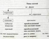
BONE STRUCTURE Every bone (lat. Os - bone) is an independent body. It has a certain shape, size, structure. Bone as an organ in an adult animal consists of the following components closely related to each other: 1) Periosteum - periosteum, located on the surface of the bone and consists of two layers. The outer (fibrous) layer is built of dense connective tissue and performs a protective function, strengthens the bone and increases its elastic properties. The inner (osteogenic) layer of the periosteum is built of loose connective tissue, in which there are nerves, vessels, and a significant number of osteoblasts (osteo-forming cells). Due to this layer, development, thickness growth and bone regeneration after injury occurs. The periosteum firmly grows together with the bone by means of connective tissue-piercing (Sharpey) fibers penetrating deep into the bone. Thus, the periosteum performs protective, trophic and osteogenic functions. A bone without a periosteum, like a tree without bark, cannot exist. The periosteum, with a bone carefully extracted from it, can re-form a bone due to the intact cells of its inner layer. 2) Compact (dense) substancesbones – substantia compacta - is located behind the periosteum and is built of lamellar bone tissue, which forms bone crossbars (beams). A distinctive feature of a compact substance is dense arrangement of bone crossbars. The strength of the compacts is provided by a layered structure and channels, inside which are vessels carrying blood. The strength of a compact substance is equivalent to cast iron or granite. 3) Spongy substance The bones - the substantia spongiosa - are located under the compact substance inside the bone and are also constructed from lamellar bone tissue. A distinctive feature of the spongy substance is that the bone crossbars are arranged loosely and form cells, so the spongy substance really resembles a sponge in structure. Compared to compact, it has much more pronounced deformation properties and is formed precisely in those places where compression and tension forces act on the bone. The direction of the bone beams of the spongy substance corresponds to the main lines of stress. Elastic deformations in the spongy substance are much stronger (4-6 times). The distribution of compact and spongy substances depends on the functional conditions of the bone. The compact substance is in those bones and in those parts of them that perform the functions of support and movement (for example, in the diaphysis of the tubular bones). In places where a large volume is required to maintain lightness and strength at the same time, a spongy substance is formed (for example, in the epiphysis of the tubular bones). 4) Inside the bone is located bone marrow cavity - cavum medullae, the walls of which are inside, as well as the surface of the bone beams are covered with a thin fibrous connective tissue sheath endosteum -endoosteum. Like the periosteum, the endosteum has osteoblasts in its composition, due to which the bone grows from the inside and recovers from fractures. 5) In the cells of the spongy substance and the bone marrow cavity is red bone marrow - medulla ossium rubra, in which the processes of blood formation. In fetuses and newborns, all the bones of the blood, but with age, gradually, the myeloid (hematopoietic) tissue is replaced with fat and red inert brain turns yellow - medulla ossium flava - and loses the function of blood formation (in domestic animals, this process starts from the second month after birth). The ratio between red and yellow bone marrow in monthly calves is 9: 1, and in adults - 1: 1. The longest red bone marrow remains in the spongy substance of the vertebrae and the chest bone. 6) Articular carp - cartilago articularis - covers the articular surfaces of the bone and is constructed from hyaline cartilage. The thickness of the cartilage varies greatly. As a rule, it is thinner in the proximal bone than in the distal one. Articular cartilage has no perchondrium and never undergoes ossification. With a large static load, it becomes thinner. Thus, in the bones of an adult animal are isolated in layers: 1) periosteum, 2) compact substance, 3) spongy substance, 4) bone marrow cavity with endosteum, 5) bone marrow, 6) articular cartilage. In the growing bone, in addition to the above 6 components, there are also other, forming bone growth zones. In such a bone there is still metaphysial cartilage, separating the body of the bone (diaphysis) from its ends (epiphyses), and three types of specially built bone tissue in contact with this cartilage and called subchondral bone. CLASSIFICATION OF BONES The classification is based on the form (structure), development and function of bones. The shape of the following types of bones: 1) Long bone(os longum) are arcuate (ribs) and tubular. They are characterized by the predominance of length over width and thickness. The tubular bones perform the function of levers of movement in the skeleton, and large amplitude movements are performed here. They distinguish the elongated part - the body, or diaphysis, and the thick ends - the epiphyses. They got their name due to the fact that a cavity for the bone marrow is formed in the middle part of the diaphysis. Between the diaphysis and the epiphysis there is a metaphysis, which, as mentioned above, ensures the growth of bones in length due to metaphysial cartilage. Among the tubular bones, there are: long tubular (shoulder, femur, forearm and lower leg bones) and short tubular (metacarpus, metatarsus, phalanxes of the fingers). It should be noted that the growth of individual bones of the skeleton can occur asynchronously. For example, the radial bone grows faster than the elbow (age deviation that does not go beyond the limits of the norm). 2) Short (spongy) bone(os breve) consist of a spongy substance, covered outside with a thin layer of compacts or articular cartilage. They have the shape of an irregular cube or polyhedron; their length, width and thickness are close in size. These include bones of the wrist and tarsus. They are located in places where high mobility is combined with a large load, and more often perform a spring function. This type of bones should also be attributed sesamoid bones, developing due to the ossification of the tendons of the muscles. 3) Flat bone (os planum ) participate in the formation of the walls of the cavities and girdles of the extremities, performing a protective function (bones of the skull roof, sternum, scapula, pelvic bones). These bones are vast surfaces for attaching muscles, they distinguish edges and corners. They consist of two layers of compacts, between which there is a small amount of spongy substance. four) Mixed bones (os irregulare, mixtum) . Have a complex shape and combine the features of the device of several types. These bones consist of several parts with different structures, outlines and origins. These include, for example, the vertebrae, the bones of the base of the skull. In some bones of the skull, a large number of veins pass, then these bones are called “diploe”. five) Pneumatic bone (os pneumaticum) have a cavity in their body (sinus, sinus) lined with mucous membrane and filled with air (maxillary, frontal, wedge-shaped). The latter may be associated with the nasal cavity. By origin distinguish : 1) Primary bones - these are bones that develop from the mesenchyme and go through only two stages of development: connective tissue and bone. These include the epithelial bones of the skull: incisal, maxillary, nasal, frontal, parietal, mezhtemennaya, scales of the temporal bone. They are characterized by endesmal (en - in, desma - connective tissue) ossification. In newborns and young animals, the integumentary bones are connected with each other and with other bones by connective tissue plates - springs (fronto-parietal, occipital-parietal). Springflows provide plasticity of the skull, which is important at birth and the growth of the skull. Primary bones also include the clavicle, the lower jaw, the proboscis bone of the pig, the sesamoid bones and the bones of the predatory penis. 2) Secondary bone - These are bones that develop from the sclerotome of the mesoderm and undergo three stages of development (connective tissue, cartilage, bone). These include most of the bones of the inner skeleton. Ossification of secondary bones occurs more difficult. Ossification, in particular, in tubular bones proceeds from three points of ossification - two epiphyseal and one diaphyseal (main points of ossification). The process of bone formation on the basis of cartilaginous buds proceeds as follows. Substitution of bone cartilage tissue includes perichondral and enchondral ossification. Perichondral ossification begins with the appearance of osteoblasts on the inner side of the perchondrium in the middle part of the diaphysis, forming a fibrous bone tissue in the shape of a cuff and then lamellar. The cartilage cells inside the perichondral girdle dissolve, the main substance of cartilage is calcified, the strength of the diaphysis increases. In this place the perichondrium becomes the periosteum, forming a bone cuff, and the perichondral ossification turns into periosteal. The formation of a bone cuff violates the nutrition of cartilage, an irreversible process of destruction begins, which is enhanced by the activity of special cells - chondroclasts. Blood vessels grow into the cavities, and along with them the elements of osteoblastic tissue penetrate, the latter is differentiated into osteoblasts and osteoclasts. Osteoclasts destroy cartilage, and osteoblasts multiply and turn into bone cells, enchondral bone occurs. Further, the periosteal and enchondral bones grow in parallel. The periosteal bone cuff grows in length to the cartilage epiphyses and in thickness. The epiphyses remain cartilaginous for some time, therefore they grow faster than the diaphysis both in length and in width. Enchondral centers of ossification appear in the epiphyses of the long bones at different times. In these centers, the calcification of cartilage occurs, its resorption, is formed first enchondral, and then perichondral bone. By the end of the fetal period in the bones may appear and additional points of ossification - apophysis, appear where the bones have significant projections, bumps. Ossified diaphysis and epiphyses join in tubular bones with cartilaginous plates - metaphysial cartilages - growth zones. Due to the metaphysical cartilage, bone growth occurs in length, with their ossification, bone growth stops. Bone growth ends when all major and additional points of ossification merge into one common bone mass.i.e. synostosis occurs. GENERAL REGULARITIES OF FORMATION OF BONES Founder of functional anatomy P.F. Lesgaftformulated a number of common patterns of bone formation. Among them, it is advisable to highlight the following: 1) Bone tissue is formedin places of greatest compression or tension; 2) The degree of bone developmentproportional to the intensity of the activity of the associated muscles. The external shape of the bones changes under the influence of tension and pressure, and the bones develop the better, the more intense the activity of the muscles associated with them. The shape and relief of the bones depends on the nature of the attachment of the muscles. So, if the muscle is attached to the bone with the help of tendons, then in this area a bump is formed, a process, and if the muscle is woven into the periosteum with a wide layer, then a depression is formed; 3) tubular and arched bone structureprovides the greatest durability and ease at the minimum expense of bone material ; 4) the external shape of the bonesdepends on the pressure on them surrounding tissues and organs and changes with decreasing or increasing pressure. The shape and position of the bones are influenced by the organs for which they form bone receptacles, pits, etc. In the places of passage of blood vessels on the bones there are necessarily grooves; 5) the restructuring of the bone shape occurs under the influence of external (for the bone) forces. The relief of bones is pronounced in old working animals and smoothed in young animals.
The bones of the embryos are made mostly of cartilage. The ossification process uses calcium to build bones as the baby grows and matures. The bones gradually become hard and strong. With age, bones lose their density and strength. When it is hard, it is called osteoporosis. Regular intake of calcium-containing foods and workouts helps bones grow and stay strong longer.
Thorax formed by the thoracic vertebrae, twelve pairs of ribs and the chest bone - sternum.
The composition of the long bones. Support - bones make up the structure for the body and provide muscle attachment points. Protection - Protection internal organs - i.e. brain, heart and lungs, help in movement. Mineral homeostasis — bone — is a repository for the production of calcium and phosphorus.
- It gives strength to hollow bone areas.
- Periosteum is a protective layer in which there is no hyaline cartilage.
- The production of blood cells occurs in the bone marrow.
Popular
- Why seals may appear
- Blood disorders in children
- What can not drink a pill
- Blood disorders in children
- Blood disorders in children
- Bones: structure, composition, types of bones, types of compounds and their characteristics
- How many live with lung cancer
- Influenza in children: how to treat, what can and cannot be done to parents, what medicines will help?
- "They are people too ..." - you say
- What helps "Prednisolone"