What bones are protected organs. What organs protects the spine? What protects the spine
The human spine, in contrast to animals, plays a huge role in the support function. In other mammals that walk on four limbs, this large and complex complex of joints is only a link for the paws. The human body, acquiring the skill of an upright position of the body, began to need an analogue of the forelimbs. In order not to lose stability, the spinal column became much larger, and also acquired physiological curves.
Vertebral infections usually develop gradually, causing constant back pain and pain to the touch. Pain worsens with movement and is not relieved by rest, heat, or analgesic use. Fever, as a rule, the most obvious sign of infection, is often absent.
When osteomyelitis is caused by a nearby soft tissue infection or direct invasion of the body, the area above the bone swells and becomes painful. An abscess may form in the surrounding tissue. These infections can not cause fever. Infection around a limb or an infected artificial limb usually causes constant pain in the region.
But due to such evolutionary changes, a person is faced with diseases of the spine, which arise from prolonged axial loads. To avoid them, nature has “equipped” the spinal column with a multitude of ligaments and muscles. Therefore, the soft tissues of the back serve not only as a support, but also as a protection for vertebral bones. They soften all the shocks and blows that inevitably form during movements.
Chronic osteomyelitis can develop if osteomyelitis is not treated successfully. This is a persistent infection that is very difficult to eliminate. Sometimes chronic osteomyelitis is not detected for a long time and does not cause symptoms for several months or years. Most often, chronic osteomyelitis causes pain in the bones, recurring infections in the soft tissues above the bone, and permanent or intermittent drainage of pus through the skin. This occurs when passage from the infected bone forms on the surface of the skin, and pus passes through it.
This complex of various structures stretched from head to toe, forming the entire back of a person along with soft tissues. Such an extended location provided protection to many internal organs. From the back they are covered with a thick layer of bones, muscles and ligaments. Therefore, diseases of the spinal column occur so often - its role in the body is too responsible.
The role of the spine in the body
Symptoms and results of an objective examination may indicate osteomyelitis. For example, doctors may suspect osteomyelitis in a subject with constant bone pain, with or without fever, and are almost always tired. If osteomyelitis is suspected, doctors perform a blood test to look for inflammation, measure the subject's red blood cell level or C-reactive protein level. In addition, blood tests often indicate a high white blood cell count. However, these blood tests are not sufficient for the diagnosis of osteomyelitis, although normal results indicating that inflammation is absent or bad, make osteomyelitis less likely.
The structure of the spine

What parts are isolated in the spinal column? For many people, it is represented by separate bones, which in the process of movement slide relative to each other. In part, this definition is correct - the joints between the vertebrae really do not communicate with each other. But in terms of anatomy, they are part of a system of complex compounds that cannot work separately. Therefore, the normal functioning of the spine is carried out only by the joint work of all vertebrae.
What protects the lumbosacral region?
X-rays can reveal characteristic changes in osteomyelitis, but sometimes not earlier than 2-4 weeks after the onset of the first symptoms. Even computed tomography and magnetic resonance imaging can identify the infected area and detect abscesses. The infected area almost always changes to scintigraphy, with the exception of children, as scintigraphy does not reliably indicate abnormal bone development. However, scintigraphy can not always distinguish infections from other bone disorders.
This anatomical formation consists of several departments that have characteristic features. Their main difference is the change in the shape and size of the vertebrae:
- The cervical region is formed by small bones that have good mobility. The absence of large processes allows them to make movements in any plane. This is due to the flexibility of the neck, which quickly turns the head in the right direction.
- The thoracic region becomes (together with the sternum) the supporting point for the attachment of the ribs. Together with these bones, they form the chest, ensuring its stability and mobility.
- The lumbar spine is formed by the largest vertebrae, as it assumes the weight of the body. But, like the neck, the bones do not have large processes, and are not fixed by other formations. Therefore, in the lower back, you can make various movements.
- The sacrum is represented by several accrete vertebrae, so it looks like a solid bone. It serves as the main support for the back, and also forms the back wall of the pelvis.
- The tailbone consists of several small bones - it was inherited by man from his ancestors, animals. It is part of the tail, which man did not need in the process of evolution.
The neural tube, which will then become part of the spine, is the first organ germ in a child growing in the womb.
How to forget about joint pain?
Scintigraphy with marked white blood cells can help distinguish between infection and other disorders in areas that look normal with bone scintigraphy. To diagnose a skin infection and identify the body that causes it, doctors can take blood samples, pus, joint fluid, or the bone itself to examine them. As a rule, osteomyelitis of the vertebrae is performed with bone needle samples or needle surgery.
The prognosis for patients with osteomyelitis is usually favorable if the treatment is timely and correct. Sometimes, however, chronic osteomyelitis develops, and bone abnormalities can recur after a few weeks, months, or even years. For children and adults who have recently developed bone infections through the bloodstream, antibiotics are the most effective method treatment. Depending on the severity of the infection, antibiotics can be administered into the vein for about 4–8 weeks, but also after oral administration.
The role of the spine in the body

This huge bone structure, willy-nilly, assumes the role of the skeleton base. She shares it with the bone pelvis, but, unlike him, she has to participate in all human movements. In addition to the support function, the spine has a number of various properties, without which the body would have a hard time:
Some people suffer from chronic osteomyelitis and need antibiotic therapy for several months. If a fungal infection identified or suspected, antimycotic drugs are administered for several months. If the infection is detected on early stage, surgery is usually not required.
For adults with bacterial osteomyelitis of the vertebrae, the usual treatment is the administration of antibiotics for 4–8 weeks. Sometimes you need to rest in bed, and the subject may have to wear a corset. Surgery may be required to remove abscesses or to stabilize the affected vertebrae.
- It interconnects the rest of the skeleton - the skull, chest, upper belt, and lower limbs. From such a peculiarity, it is not easy for him - he has to participate in any movement.
- Cartilaginous intervertebral discs and ligaments play the role of a shock absorber, softening any human movement. If they were not there, then at every step people would shake from head to toe.
- It provides a vertical position of the body - it may seem that when standing, the spine is at rest. But this is not so - in this position almost all the muscles of the back are strained.
- Physiological curves - lordosis and kyphosis - do not allow soft tissues to tire easily, as they evenly distribute the load between them.
- The spinal column is a place where many soft tissues adhere. Muscles, tendons, vessels, nerves - all this passes along or through it. And it is so arranged that it does not allow even minimal damage to them.
Due to the large length of the spine, it is in contact in each department with different organs, which makes such a complex structure of the back.
What protects the spine?
In addition to various organ systems, this complex joint contains its own formations that must be protected. For them in the bodies of the vertebrae there are many holes through which they freely pass. But the main formation that stretches across the entire pillar is the spinal canal containing the spinal cord:
- This organ is protected from external influences by the vertebrae themselves, as well as by a two-layer sheath. They produce a soft and hard layer that perform opposite functions.
- When moving, the spinal cord does not stretch, as it is located freely relative to the shells.
- Its whole tissue is located only at the level of the cervical and thoracic - in the lumbar region it already consists of many small nerves.
- Inside the soft shell circulates cerebrospinal fluidwhich protects the body from tremors. It also provides its nutrition and exchange products.
- The vertebrae are interconnected by a whole capsule, which eliminates the contact of the spinal cord with other organs during movements.
This system of protection is due to the role of the spinal cord - it ensures the operation of all parts of the body and internal organs.
What protects the cervical?

The neck is not only a moving part of the body, but also contains all the paths connecting the head and torso. Therefore, the greatest number of structures is observed precisely in the cervical region, where the spine is surrounded by many vessels and nerves:
- The vertebrae contain in their canal a part of the spinal cord, which is responsible for the functioning of the vital organs - the heart and lungs.
- The vertebral arteries, which are the main source of nutrition for the brain (together with the carotid arteries), pass through their processes.
- Bones provide support for neck organs - respiratory tract, esophagus. Back wall the pharynx is completely attached to the vertebrae. If it were not for such protection, the person could not breathe freely and swallow.
- The vertebrae provide the normal position of the vascular bundles that provide blood supply to the brain.
- In this section are located the nerve plexus, which ensures the functioning of the upper limbs.
Therefore, neck injuries are so dangerous - even a slight dislocation can damage important structures.
What protects the thoracic?

The thoracic vertebrae form the basis for the chest - a solid framework of ribs closes on them. This firm and flexible structure closes all organs located in the chest:
- Together with the pectoral muscles, respiratory movements are created that ensure the mobility of the lungs. Inhalation and exhalation are carried out due to alternate changes in the tone of these muscles.
- The heart is firmly hidden behind the bony structures, like a stone wall.
- A number of important vessels pass along the thoracic segment - the aorta, the inferior vena cava, the main lymphatic trunk. They are located on the side of the spine, and secured with soft tissues. Their damage is fatal to humans, so they are covered behind a layer of bones, muscles and ligaments.
- The tissue of the liver and spleen is easily affected and bleeds in the event of injury, so it is safely hidden from them behind the bone structures. Their shell is connected to the vertebrae by a multitude of ligaments, which create their immobility.
The thoracic region is treated as one with the ribs and sternum, as it is firmly connected with the joints.
What protects the lumbosacral region?

This part of the back contains a powerful layer of muscles that support the human body in an upright position. It also provides protection to many organs and entities located in this segment:
- The kidneys are entirely dependent on the lumbar vertebrae - their soft tissues form a special “bed” for them. It is a pocket in which they are without the support of ligaments. A large thickness of the muscles keeps them warm, which is vital for them.
- Aorta and large veins run in the direction of the lower limbs along the entire loin. Large vertebrae protect them from damage from the back.
- The intestine is in an orderly position, as the folds attached to the anterior surface of the vertebrae are directed to its loops.
- The lumbar region and the sacrum serve as a receptacle in which powerful nerve plexuses are located. They form a multitude of nerves heading towards the lower limbs. Here also the protection of the spinal cord is performed, since these fibers are its final divisions.
Together with such "responsibility" the loin should have sufficient mobility. Therefore, all the formations are located at a sufficient distance so that during the movements they would not be infringed.
How to forget about joint pain?
- Joint pains limit your movement and full life ...
- You are worried about discomfort, crunch and systematic pain ...
- Perhaps you have tried a bunch of drugs, creams and ointments ...
- But judging by the fact that you are reading these lines - they did not help you much ...
But orthopedist Sergei Bubnovsky claims that indeed effective remedy from joint pain exists!
Work 2.
1. Read § 1 “Human Sciences”, fill in the table.
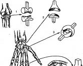
2. Consider in the textbook a reproduction of Rembrandt’s “Anatomy Lesson of Dr. Tülp” picture (Fig. 1) and in the “Functions” column describe the work of the forearm muscles.
Structure.
The muscles are located on the palmar side of the forearm, have a tape-like shape. At one end they are attached to the bones of the forearm, the other to the bones of the fingers.
Functions.
Finger flexion.
Work 3.
Read the article “Body Structure. The place of man in nature ”(§ 2). Determine the ratio of body parts.
1. Measure and compare the length of the ear (6 cm) and nose (6 cm); the length of the forearm (22 cm) and feet (22 cm).
2. Measure the knee circumference (29 cm). Does the knee circumference really fit the neck circumference?
The circumference of the neck is equal to the circumference of the knee.
(Figures may be different for boys and girls!).
Work 4.
Consider drawing “ Internal organs of man "on the first flyleaf of the textbook.
1. Locate the thoracic obstruction, organs of the thoracic and abdominal cavities. Write down the names of these bodies and put the numbers by filling out the table.

2. Fill in the blanks in the text.
The right lung has three lobes. The liver is located in the abdominal cavity right upper quadrant, stomach - in the abdominal cavity. The appendix is in the right groin.
Work 5.
1. Insert the missing words.
In the cranial cavity are the cerebral hemispheres, the diencephalon, the midbrain, the cerebellum, the medulla, the bridge, and the cerebral vessels.
In the cavity of the spinal canal is the spinal cord.
In the chest cavity are the heart, blood vessels and nerves, lungs, respiratory tract.
In the abdominal cavity are the stomach, liver, spleen, intestines thick and thin, the pancreas, kidneys, blood vessels and nerves.
2. List the organs that begin in the chest cavity, and end in the abdominal cavity.
Esophagus, vessels and nerves.
3. What bones protect the abdominal organs?
The spine is behind, pelvic bones - from below.
4. Take a deep breath and exhale and follow the movements of the abdominal wall. Could its wall move easily if it had a bone base?
Could not. The abdominal wall does not have a bone basis, but only the abdominal muscles. This ensures the normal breathing process: inhale and exhale, the work of the diaphragm and chest muscles.
5. Look at the table “Man in the system of the organic world” (A) and write down the biological signs that allow
feed the young milk: the presence of mammary glands in females, nasal and oral cavity separated by a soft and hard palate, there is no gap between the teeth and cheeks;
maintain a constant body temperature: hair and subcutaneous fatty tissue;
maintain a high metabolism due to the peculiarities of the circulatory system: the heart is four-chamber, completely divided into 2 atria and 2 ventricles, one aorta extends from the heart;
respiratory system: lungs of the alveolar type, the air in them enters the tubes forming the bronchial tree.
6. Review the list of features that make it possible to assign a person to the primate order (Table 1, B), and write down the signs that allow
grab items: five-fingered limbs, the thumb is opposed to the rest, fingers and toes are provided with flat nails, not claws;
develop mental activity: complication of the anterior part of the brain, in particular, the large hemispheres.
7. Write down from the table. 1 (B) morphological features that allow higher primates and humans
increase the speed of movement: short body and long limbs;
go to erect: reduction of the caudal vertebrae, decrease in the number of thoracic and lumbar vertebrae, increase in the number of sacral vertebrae fused into the sacrum.
8. Write down from the table. 1 (D) morphological features that allowed a person to develop sound speech: brain department the skull is larger than the facial one, the canines are equal to the incisors, there is a mental protrusion to which the muscles responsible for speech are attached, the maxillary part of the skull does not come forward;
to adapt to work: the sternum is articulated with the clavicle, the radius is movable relative to the ulna, the bones of the palm are movable, the thumb is contrasted with the rest;
to master an upright movement: the attachment of the skull to the spine almost coincides with the center of gravity of the head, the spine is S-shaped, rib cage flattened in the dorso-ventral direction, the bones of the pelvis and lower extremities are massive, the foot is vaulted, the toe is shortened, the thumb is not contrasted with the rest;
to lead a social life: the development of the cerebral cortex.
Popular
- How many live with lung cancer
- Influenza in children: how to treat, what can and cannot be done to parents, what medicines will help?
- "They are people too ..." - you say
- What helps "Prednisolone"
- How are menstruation for endometriosis?
- The fourth stage of cancer, how many live with it
- Prednisolone ampoules instructions for use
- Effective treatments for inflammation of the appendages
- Sinupret - an effective herbal alternative to antibiotics in the treatment of sinusitis
- When do menstruation begin for the first time