The structure of the bones of the pelvic girdle. Bones of the free lower limb. Anatomy of the joints of the human lower limbs. Sacroiliac joint, pubic symphysis
Introduction
Legs or lower limbs bear the brunt of the burden, because they have to perform the functions of support and movement. These are the most powerful of all the muscles of the human body.
The lower limbs are divided into the muscles of the pelvis and the muscles of the free lower limb. The latter group is divided into the muscles of the thigh, lower leg and foot. The pelvic muscles originate from the bones of the pelvis, sacrum and lumbar vertebrae, and also attach to the femur. Their function is to hold the torso in an upright position, flexion, extension in the hip joint and movement of the hip. The muscles of the thigh are divided into three groups: anterior, posterior and medial (leading). These muscles are also involved in keeping the body upright, but their main role is movement. The muscles of the leg are also divided into three groups: anterior, posterior and lateral (lateral). They are responsible for dorsal flexion and plantar extension of the foot, the support functions of the foot and its orientation. The muscles of the foot are similar to the muscles of the hand, divided into 3 groups. In connection with the upright man, they have a special purpose. The main function of the muscles of the foot - ensuring stability in various positions of the foot, flexion, extension and abduction of the fingers.
The purpose of this work is to study the features of the structure of the skeleton, the muscles of the lower limb, their blood supply and innervation.
1. The structure of the bones of the pelvic girdle and free lower limb
Pelvic bone, os plow, (see figure) as a whole bone is present in adults. Up to 14-16 years old, this bone consists of three separate bones connected by cartilage: the iliac, pubic and sciatic. The bodies of these bones on their outer surface form the acetabulum, acetabulum, which is the articular fossa for the femoral head. The acetabulum is deep, circumferentially circumscribed by a high margin, which on its medial side is interrupted by cutting the acetabulum, incisura acetabuli. For articulation with the femoral head in the acetabulum there is a semi-lunar surface, facies lunata, which occupies the peripheral part of the acetabulum. The center of the acetabulum is the fossa of the acetabuli, fossa acetabuli, - rough and somewhat indented. The ileum, os ilium (ilii), consists of two sections: the lower thickened section - the body of the ileum, corpus ossis ilii, participates in the formation of the acetabulum; the upper, extended section - the iliac wing, ala ossis ilii. The iliac wing is a broad curved plate, thinned in the center. To the periphery, the bone plate thickens, expanding fan-like upward, and ends with a convex edge - the iliac crest, crista iliaca. On the iliac crest, there are three rough lines for attaching the wide abdominal muscles: the outer lip, labium externum, inner lip, labium internum, and the intermediate line, linea intermedia. The iliac crest in front and behind has bony protrusions - the upper and lower iliac spines.
Anterior superior iliac spine, spina iliaca anterior superior. Below it, separated by tenderloin, is the lower anterior iliac spine, spina iliaca anterior inferior. At the posterior end of the crest, a protrusion is visible - the upper posterior iliac spine, spina iliaca posterior superior, and slightly below it, the lower posterior iliac spine, spina iliaca posterior interior. On the outer surface of the wing of the Ilium, there are weakly defined three rough lines. Of these, the anterior gluteal line, linea glutealis anterior, is more visible. It is the longest, starts from the upper anterior iliac spine, goes arcuate toward the large sciatic notch of the ischium. The posterior gluteal line, linea glutealis posterior, is much shorter, located below the previous one and is oriented almost vertically. The lower gluteal line, linea glutealis inferior, shorter than other lines, begins between the upper and anterior inferior iliac spine and goes along a gentle arc over the acetabulum to a large sciatic notch. On the inner concave smooth surface of the wing of the Ilium there is a gentle depression - the iliac fossa, fossa iliaca. The lower border of the iliac fossa is the arcuate line, linea arcuata. The beginning of this line is the ear-shaped surface, facies auricularis, its leading edge. This surface is used for articulation with the same surface of the sacrum. The arcuate line extends anteriorly to the iliac-pubic eminence. Above the uviform surface is the iliac tuberosity, tuberositas iliaca, for the attachment of the interosseous ligaments. The pubis, os pubis, has an expanded part - the body and two branches. The body of the pubic bone, corpus ossis pubis, forms the anterior section of the acetabulum. An upper branch of the pubic bone, ramus superior ossis pubis, bearing the ileal-pubic eminence, eminentia iliopubica, located along the fusion line of the pubic bone with the ileum, is sent forward. The front of the upper branch sharply curves downwards and is regarded as the lower branch of the pubic bone, ramus inferior ossis pubis. In the place where the upper branch passes into the lower, in the medial region there is an oval symphysial surface, facies symphysialis, which serves to connect with the pubic bone of the opposite side. On the upper branch of the pubis, about 2 cm from the medial end, there is a pubic tubercle, tuberculum pubicum, from which the pubic crest, crista pubica, extends laterally to the posterior ileal pubic elevation on the lower surface of the upper branch. pubic bone. In the direction backwards and medially the locking groove, sulcus obturatorius, passes for the vessels of the same name and nerve.
The ischium, os ischii (ischium), has a thickened body, corpus ossis ischii, which supplements the acetabulum from below and passes into the branch of the ischial bone, ramus ossis ischii. The body of the sciatic bone is with the branch angle, open to the front. In the corner of the bone forms a thickening - sciatic tuber, tuber ischiadicum. Above this mound, the sciatic spine, spina ischiadica, separates two cuts from the rear edge of the body: the lower - small sciatic notch, incisura ischiadica minor, and the large sciatic notch, incisura ischiadica major, located at the level of the upper edge of the acetabulum. The branch of the sciatic bone is connected to the lower branch of the pubic bone, thus closing the oval obturator opening, the foramen obturatum, the pelvic bone, from below.
Skeleton of the free part of the lower limb
The femur, femur (os femoris), is the largest and longest tubular bone in the human body. (fig. 066) Like all long tubular bones, it has a body and two ends; at the upper proximal end is located the head of the femur, caput ossis femoris, for connection with pelvic bone. The articular surface of the head is directed medially and upwards. In the middle of it is the fossa of the femoral head, fovea capitis femoris, - the place of attachment of the femoral head ligament. The femoral neck, соllum femoris, connects the head with the body and forms an angle of about 130 ° with it. On the border of the neck and body, there are two powerful bony tubercles called spits. The greater trochanter, the trochanter major, is located at the top and laterally, on its medial surface facing the neck, there is a spitting fossa, fossa trochanterica. The trochanter minor spit is located at the lower edge of the neck, medially and posteriorly. In front, both skewers are interconnected by an intertrochanter line, linea intertrochanterica, from the rear, an intertrochanic crest, crista intertrochanterica.
The body of the femur, corpus femoris, is approximately cylindrical in shape, curved convexly anteriorly and as if twisted around its longitudinal axis. The body of the bone is smooth, on the back surface there is a rough line, linea aspera, which is divided into the medial and lateral lips, labium mediale et labium laterale. In the middle of the femur, the lips closely adjoin one another, and they diverge towards the top and bottom. Up lips go to the big and small spits of the femur. The lateral lip significantly expands and thickens, passing the gluteal tuberosity, tuberositas glutea, - the place of attachment of the gluteus maximus muscle. Sometimes gluteal tuberosity acts as a third spit, trochanter tertius. The medial lip extends into a rough comb line, linea pectinea. At the lower end of the femur, both lips gradually move apart from one another, limiting the triangular shape of the popliteal surface, facies poplitea.
The distal end of the femur is enlarged and forms two large rounded condyle, which differ in size and degree of curvature of the articular surfaces. The medial condyle, condylus medialis, is larger than the lateral condylus lateralis. They are located on the same level, since the femur in its natural position is tilted so that its lower end is closer to the median line than the upper one. Both condyles are separated from each other by a deep inter-cracked fossa, fossa intercondylaris. Above the articular surface of the medial condyle is the medial epicondyle, epicondylus medialis. On the lateral side of the same condyle there is a smaller lateral epicondyle, epicondylus lateralis. In front, the articular surfaces of the condyles pass into each other, forming a concave patellar surface, facies patellaris, to which the patella is attached with its back side.
The patella, patella, is a large sesamoid bone enclosed in the quadriceps tendon. The patella is flattened in the anteroposterior direction. Allocate the base of the patella, basis patellae, directed upwards, and the top of the patella facing down, apex patellae. The articular surface, the facies articularis, the patella articulates with the patellar surface of the femur, the anterior surface, the facies anterior, rough and easily palpable through the skin.
The crus, crus, consists of two bones: a medially located tibial bone and a laterally-fibe bone. Both belong to the long tubular bones; in each of them there is a body and two ends. The ends of the bones are thickened and carry surfaces to join with the femur at the top (tibia) and the bones of the foot at the bottom. Between the bones there is an interosseous space of the tibia, spatium interosseum cruris.
Tibia, tibia. In length, this bone occupies the second place in the human skeleton and is the thickest bone of the tibia. The proximal bone end is significantly thickened and has a medial and lateral condyle, condylus medialis et condylus lateralis. The upper articular surface, the facies articularis superior, faces upwards and articulates with the condyles of the femur. The articular surfaces of the tibial condyles are separated by an inter-muscular elevation, eminentia intercondylaris, which consists of two tubercles: the medial inter-muscular tubercle, tuberculum intercondylare mediale, and the lateral inter-muscular tubercle, tuberculum intercondylare laterale. Ahead of the inter-muscular elevation is the anterior inter-mussel field, area intercondylaris anterior, behind the posterior inter-muscular field, area intercondylaris posterior. Below the lateral condyle with its lateral side and somewhat posteriorly, there is the fibular articular surface, facies articularis fibularis, for articulation with the fibula.
The body of the tibia, corpus tibiae, triangular. The anterior margin, margo anterior, is the sharpest, is palpable through the skin, thickens at the top and forms the tibial tuberosity, tuberositas tibiae, to which the quadriceps muscle is attached. The lateral margin is also sharp and faces the fibula, so it is known as the interosseous margin, margo interosseus. The medial margin, margo medialis, is somewhat rounded. In addition to the edges, there are three surfaces in the body of the tibia. The medial surface, the facies medialis, is smooth, lying directly under the skin. The lateral surface, the facies lateralis, and the posterior surface, the facies posterior, are covered with muscles. On the posterior surface of the bone, the rough line of the soleus muscle is visible, the linea musculi solei, which runs from the posterior edge of the lateral condyle downward and medially; here begins the muscle of the same name. Below this line is a large feeding hole leading to a canal distally directed.
The distal end of the tibia is enlarged and approximately quadrangular in shape. On the lateral edge of the distal end of the tibia is the fibular incision, incisura fibularis, for connection with the fibula. On the medial side of the tibia, the medial ankle, malleolus medialis, extends downwards. Behind it is a shallow ankle groove, sulcus malleolaris, for the posterior tibialis muscle tendon here. On the lateral side of the medial ankle is the articular surface of the ankle, facies articularis malleoli, which at an angle passes into the lower articular surface, facies articularis inferior, of the tibia. These surfaces, together with the articular surface of the fibula, are articulated with the talus bone of the tarsus (foot).
Fibula, fibula. This bone is much thinner than the tibial and almost the same length as it. At the proximal thickened end is the head of the fibula, caput fibulae. It highlights the upward tip of the head of the fibula, apex capitis fibulae, and on the medial side is the articular surface of the head of the fibula, facies articularis capitis fibulae, for articulation with the tibia. The head is narrowed downwards and with the help of the fibular neck, collum fibulae, passes into the bone body.
The body of the fibula, corpus fibulae, is trihedral, somewhat twisted along its longitudinal axis, slightly curved in the upper part in the medial direction. The body distinguishes the anterior margin, margo anterior, the posterior margin, margo posterior, and the medial sharp interosseous margin, margo interosseus. Three surfaces are limited to these edges: the lateral surface, the facies lateralis, the posterior surface, the facies posterior, and the medial surface, the facies medialis.
The lower distal end of the fibula is thickened and forms the lateral ankle, malleolus lateralis, which is longer than the medial ankle of the tibia. On the medial surface of the lateral ankle, the smooth articular surface of the ankle, facies articularis malleoli, stands out, to connect with the talus. Behind the articular surface of the ankle is the fossa of the lateral ankle, fossa malleoli lateralis, to which the tendons of the peroneal muscles are attached.
The bones of the foot, ossa pedis, as well as the bones of the hand, are divided into three sections: bones of the tarsus, ossa tarsi, metatarsal bones, ossa metatarsi, and bones of the fingers (phalanges), ossa digitorum (phalanges) (Fig. 068).
The bones of the tarsus, ossa tarsi, include seven spongy bones arranged in two rows. The proximal (posterior) row consists of two large bones: ankle and heel; the remaining five bones of the tarsus form a distal (anterior) row.
The talus, talus, has a body, corpus tali, head, caput tali, and a narrow part connecting them - the neck, collum tali. The body of the talus is the largest part of the bone. Its upper part is a block of the talus, trochlea tali, with three articular surfaces. The upper surface, facies superior, is intended for articulation with the lower articular surface of the tibia.
Two other articular surfaces lying on the sides of the block: the medial ankle surface, the facies malleolaris medialis, and the lateral ankle surface, the facies malleolaris lateralis, are articulated with the corresponding articular surfaces of the tibial and fibula bones. The lateral ankle surface is much larger than the medial and reaches the lateral process of the talus, processus lateralis tali.
Behind the block, the posterior process of the talus, processus posterior tali, departs from the body of the talus. The tendon groove of the long flexor of the toe, sulcus tendinis musculi flexoris hallucis longi, divides this process into the medial tubercle, tuberculum mediale, and the lateral 6 ovule, tuberculum laterale. On the underside of the talus there are three articular surfaces for articulation with the calcaneus: the anterior calcaneal articular surface, facies articularis calcanea anterior; middle calcaneal articular surface, facies articularis calcanea media, and posterior calcaneal articular surface, facies articularis calcanea posterior. Between the middle and posterior articular surfaces, there is a fodder of the talus, sulcus tali. The head of the talus is directed forward and medially. To articulate it with the navicular bone, there is a rounded navicular articular surface, facies articularis navicularis.
The calcaneus, calcaneus, is the largest bone of the foot. It is located under the talus and significantly protrudes from under it. Behind the body of the calcaneus has a tubercle of the calcaneus bent downwards, tuber calcanei. On the upper side of the calcaneus, three articular surfaces are distinguished: the anterior talaric articular surface, the facies articularis talaris anterior, the average talaric articular surface, the facies articularis talaris media, and the posterior articular articular surface, facies articularis talaris posterior. These articular surfaces correspond to the calcaneal articular surfaces of the talus. Between the middle and posterior articular surfaces, there is a sulcus of the calcaneus, sulcus calcanei, which, together with the corresponding sulcus on the talus, forms the tarsal sinus, sinus tarsi, the entrance to which is located on the back of the foot from the lateral side.
From the anterior surface of the calcaneus, a short and fat process extends from the medial side - the support of the talus, sustentaculum tali. On the lateral surface of the calcaneus, the sinus furrow of the long peroneal muscle passes, sulcus tendinis musculi peronei (fibularis) longi. The distal (anterior) end of the calcaneus has a cuboid articular surface, facies articularis cuboidea, for articulation with the cuboid bone.
Scaphoid bone, os naviculare, is located medially, between the talus and three cuneiform bones. The proximal concave surface is articulated with the head of the talus. Distal surface of navicular bone more proximal; it has three articular pads for joining with the sphenoid bones. The tuberosity of the navicular bone, tuberositas ossis navicularis (place of attachment of the posterior tibial muscle) is noticeable at the medial margin. On the lateral side of the navicular bone there may be a non-permanent articular surface for articulation with the cuboid bone.
The wedge-shaped bones, ossa cuneiformia (medial, intermediate and lateral), are located anterior to the navicular bone and are located in the medial part of the foot. Of all the bones, the medial sphenoid bone, os cuneiforme mediale, is the largest, articulated with the base of the first metatarsal bone; intermediate sphenoid bone, os cuneiforme intermedium, with II metatarsal bone; lateral sphenoid bone, os cuneiforme laterale, with III metatarsal bone.
The cuboid bone, os cuboideum, is located on the lateral side of the foot between the calcaneus and the last two metatarsal bones. The joints of these bones have articular surfaces. In addition, on the medial side of the cuboid bone there is an articular platform for the lateral sphenoid bone, and a few posterior and smaller sizes - for articulation with the scaphoid bone. On the lower (plantar) side there is a tuberosity of the cuboid bone, tuberositas ossis cuboidea, in front of which is the sinus furrow of the long peroneal muscle, the sulcus tendinis musculi peronei (fibularis) longi.
The metatarsal bones, ossa metatarsi, are five tubular short bones. The shortest and thickest - I metatarsal bone, the longest - II. As in the metacarpal bones, the body of the metatarsal bone is isolated, - corpus metatarsale, head, caput metatarsale, and base, basis metatrsalis. The bodies of the metatarsal bones have a prismatic shape with a bulge facing in back side. The bases are provided with articular surfaces for articulation with tarsal bones. The head of the first metatarsal bone on the plantar side is divided into two areas, to which the sesamoid bones are attached. The base of the first metatarsal bone forms a joint with a medial sphenoid bone. The bases of the II and III bones articulate with the intermediate and lateral sphenoid bones, and the bases of the IV and V metatarsal bones - with the cuboid bone. On the lateral side of the V metatarsal bone is the tuberosity of the V metatarsal bone, tuberositas ossis metatarsalis V, for the attachment of the short peroneal muscle.
The bones of the fingers (phalanges), ossa digitorum (phalanges), and the feet differ from the bones of the fingers of the hand in their size: they are much shorter. The toes, as well as on the hand, have a proximal phalanx, phalanx proximalis, middle phalanx, phalanx media, and distal phalanx, phalanx distalis. The exception is the thumb (I finger), hallux (digitus primus), the skeleton of which consists of two phalanges: proximal and distal. The phalanges are tubular bones. There are phalanx body, corpus phalangis, phalanx head, caput phalangis, phalanx base, basis phalangis, and two ends. The bodies of the proximal and middle phalanges are somewhat convex to the back. The base of each proximal phalanx has a flattened fossa, which serves to form a joint with the head of the corresponding metatarsal bone. On the basis of the middle and distal phalanges, there are two flat pits separated by a comb for articulation with the phalanx head located more proximally. Each distal (nail) phalanx ends with a tubercle, tuberositas phalangis distalis.
The bones of the tarsus and metatarsus do not lie in the same plane. The talus is located on the heel bone, and the scaphoid bone is above the heel bone and cuboid. The bones of the medial margins of the tarsus are elevated in comparison with its lateral margin. With this interposition of the bones of the foot, its arches are formed, which provide spring support for the lower limb. The arch of the foot has a bulge facing upwards. In fact, the foot rests on the ground only at a few points: behind it is the heel of the calcaneus, in front of the head of the metatarsal bones, mainly II and V. The phalanges only touch the ground slightly.
2. Connection of the lower limb
Sacroiliac joint, articulatio sacroiliaca, paired joint, formed by the ilium and sacrum. The articular articular surfaces, facies auriculares, of the ilium and the sacrum are flat, covered with fibrous cartilage. The articular capsule, capsula articularis, attaches along the edge of the articular surfaces and is tightly stretched. The ligamentous apparatus is represented by strong, tightly stretched fibrous bundles located on the front and rear surfaces of the joint. On the front surface of the joint are ventral sacroiliac ligaments, ligg. sacroiliaca ventralia. They are short bundles of fibers that run from the pelvic surface of the sacrum to the ilium.
On the back of the joint are several ligaments:
Interosseous sacroiliac ligaments, ligg. sacroiliaca interossea, lie behind the sacroiliac joint, in the gap between the bones that form it, and attach their ends to the iliac and sacral tuberosities;
Dorsal sacroiliac ligaments, ligg. sacroiliaca dorsalia, individual bundles of these ligaments, starting from the posterior inferior spine of the Ilium, are attached to the lateral crest of the sacrum along the II-III sacral openings. Others follow from the posterior superior iliac spine downwards and somewhat medially, attaching to the posterior surface of the sacrum in the region of the IV sacral vertebra. The sacroiliac joint belongs to the sedentary joints.
The pelvic bone, in addition to the sacroiliac joint, is connected to the spinal column through a series of powerful ligaments, which include the following:
Sacro-lumpy bundle, lig. sacrotuberale, starts from the medial surface of the sciatic tubercle and, going up and medially, expands fan-shaped, attaching to the outer edge of the sacrum and tailbone. Part of the fibers of this ligament passes to the lower part of the branch of the ischium and, continuing along it, forms the sickle process, processus falciformis;
Sacrospinous ligaments, lig. sacrospinale, starts from spina ischiadica, goes medially and posteriorly and, being located in front of the previous ligament, attaches along the edge of the sacral bone and partly the coccyx. Both ligaments together with the large and small sciatic notches limit two holes: the large sciatic, foramen ischiadicum majus, and the small sciatic, foramen ischiadicum minus. Both of these openings allow muscles as well as vessels and nerves to leave the pelvis;
Ileo-lumbar ligament, lig. iliolumbale, starts from the anterior surface of the transverse processes of the IV and V lumbar vertebrae, goes outwards and attaches to the posterior parts of the crista iliaca and the medial surface of the iliac wing;
Sacrococcygeal junction, junctura sacrococcygea (see junction of the spine).
The pubic symphysis, symphysis pubica, is formed by the hyaline cartilage-covered articular surfaces of the pubic bones, the facies symphysialis, and the fibrous cartilaginous interlocal disk located between them, discus interpubicus. The specified disk grows together with the articular surfaces of the pubic bones and has a sagitally located slit-like cavity in its thickness. In women, the disc is slightly shorter than in men, but thicker and has a relatively large cavity.
The pubic symphysis is fortified with the following bundles:
Upper pubic ligament, lig. pubicum superius, which is on the upper edge of the symphysis and stretched between both tuberculum pubicum;
Arc ligament pubis, lig. arcuatum pubis, which on the lower edge of the symphysis moves from one pubic bone to another.
Locking membrane, membrana obturatoria, consists of bundles of connective tissue fibers, mainly in the transverse direction, which are attached along the edge of the foramen obturatum, performing it all the way, with the exception of sulcus obturatorius. The locking membrane has a number of small holes. She and the muscles starting from it, along with the sulcus obturatorius, restrict the obturator canal, canalis obturatorius, through which the vessels of the same name and nerve pass through it.
3. Muscles of the lower limbs
The muscles of the thigh surround the femur and are divided into an anterior muscle group, which consists mainly of the extensors, the medial group, which includes the adductors, and the posterior muscle group, including the flexors
Front group. Tailor's muscle (m. Sartorius) (Fig. 1, 2, 3, 4) flexes the thigh and shin, simultaneously rotating the thigh outwards, and the thigh inward, providing the ability to throw one's legs over the leg. It is a narrow ribbon, located on the front surface of the thigh and, spiraling down, goes to the front surface. Tailor's muscle is one of the longest muscles of a person. It starts from the upper anterior iliac spine, and is attached to the tibial tuberosity and with separate tufts on the fascia of the tibia.
|
Fig. 1. The muscles of the pelvis and thighs (front view): 1 - pear muscle; |
|
|
Fig. 2. The muscles of the pelvis and hips (side view): 1 - large lumbar muscle; |
|
|
Fig. 3. The muscles of the pelvis and hips (side view): 1 - the broadest muscle of the back; |
|
|
Fig. 4. The muscles of the pelvis and thighs (rear view): 1 - gluteus maximus; |
The quadriceps muscle of the thigh (m. Quadriceps femoris) (Fig. 1) consists of four heads and is the largest muscle of a man. With the reduction of all heads, it extends the lower leg, while reducing the rectus femoris muscle, it participates in its flexion.
Located on the anterolateral surface of the thigh, in the lower sections completely goes to the side. Each head has its own starting point. The longest rectus femoris muscle (m. Rectus femoris) (Fig. 2) begins on the inferior iliac spine; the medial broad muscle of the thigh (m. vastus medialis) (Fig. 2, 3) - on the medial lip of the rough femur line; lateral broad muscle of the thigh (m. vastus lateralis) (Fig. 1, 3) - on the greater trochanter, intertrochanteric line, and lateral lip of the rough line of the femur; the intermediate broad muscle of the thigh (m. vastus intermedius) - on the front surface of the femur. All heads grow together, forming a common tendon, which is attached to the top and side edges of the kneecap, bypassing which the tendon falls below and goes into the knee ligament, which is attached to the tibial tuberosity. In the place of attachment of the muscles, there are the patellar pouch (bursa suprapatellaris), the subcutaneous podrenal bag (bursa subcutanea prepatellaris), the subcutaneous podnatserennial bag (bursa subcutanea infrapatellaris) and the deep undergrown bag (bursa infrapatellaris profunda).
The knee joint muscle (m. Articularis genus) tightens the bag knee joint. It is a flat plate and is located on the front surface of the thigh under the intermediate broad muscle of the thigh. The point of its beginning is on the front surface of the lower third of the femur, and the attachment point is on the front and side surfaces of the articular bag of the knee joint.
Medial group. The scalloping muscle (m. Pectineus) (Fig. 2) flexes and leads the hip, rotating it outwards. The flat muscle is quadrangular in shape, starts on the ridge and upper branch of the pubic bone, and is attached on the medial lip of the rough line of the femur below the small skewer.
Thin muscle (m. Gracilis) (Fig. 2, 4) leads the thigh and takes part in flexing the tibia, turning the leg inward. The long flat muscle is located directly under the skin. The point of its beginning is on the lower branch of the pubic bone, and the place of attachment is on the tibial tuberosity. The tendon of the thin muscle grows together with the tendons of the tailoring and semi-tendinous muscles and the fascia of the tibia, forming a superficial goose foot. Here is the so-called goose bag (bursa anserina).
The long adductor muscle (m. Adductor longus) (Fig. 2) causes the thigh to participate in its flexion and rotation outwards. This is a flat muscle, having the shape of an irregular triangle and located on the anteromedial thigh. It starts from the upper branch of the pubic bone and is attached on the middle third of the medial lip of the rough line of the femur. The short adductor muscle (m. Adductor brevis) (Fig. 1) causes the thigh to participate in its flexion and rotation outwards. It is a muscle of a triangular shape, starts on the front surface of the lower branch of the pubic bone, lateral to the thin muscle, and is attached on the upper third of the medial lip of the rough line of the femur.
The large adductor muscle (m. Adductor magnus) (Fig. 1, 2, 4) causes the thigh, partly rotating it outwards. Thick, wide, the most powerful of this group of muscles, which is located deeper than the rest of the adductor muscles. The point of its beginning is on the sciatic hill, as well as on the branch of the sciatic bone and the lower branch of the pubic bone. The attachment site is located on the medial lip of a rough line and medial epicondyle of the femur. In the muscle bundles, several holes are formed which allow blood vessels. The largest of these is called the tendon orifice (hiatus tendineus). Above it is the fascial plate, and between it and the muscle a space of triangular shape is formed, called the lead channel (canalis adductorius) (Fig. 1). The femoral vein, artery and hidden nerve of the lower limb pass through it.
Posterior group The biceps femoris muscle (m. Biceps femoris) (Fig. 3, 4) extends the thigh and bends the lower leg. In the bent position rotates the shin outwards. Passes along the side edge of the upper surface of the thigh. The muscle has one abdomen and two heads. The long head (caput longum) starts from the sciatic hill, the short head (caput breve) - on the lower part of the lateral lip of the rough line of the femur. The abdomen ends with a long narrow tendon, the attachment of which is located on the head of the fibula. Part of the bundles are woven into the fascia of the leg. Near the point of the beginning of the long muscle is the upper bag of the biceps femoris (bursa m. Bicipitis femoris superior). In the area of the tendon is the lower tummymine sac of the biceps femoris (bursa subtendinea m. Bicipitis femoris inferior).
The semi-tendinous muscle (m. Semitendinosus) (Fig. 2, 4) extends the thigh, bends the lower leg, rotates it in the bent position, and also participates in the extension of the trunk. The muscle is long and thin, partly covered by the gluteus maximus, sometimes interrupted by a tendon bridge (intersectio tendinea) (Fig. 4). The point of its beginning is located on the sciatic tubercle, and the place of attachment is on the medial surface of the tibial tuberosity. Separate bundles of muscles are woven into the fascia of the leg, taking part in the formation of a goose foot.
The semi-membranous muscle (m. Semimembranosus) (Fig. 2, 4) extends the thigh and bends the lower leg, rotating it inwards. Passes along the medial edge of the posterior surface of the thigh and partially covers the semitendinosus muscle. The muscle starts from the sciatic hill and is attached at the edge of the medial condyle of the tibia.
The tendon is divided into three bundles, forming a deep goose foot. The external bundle passes into the popliteal fascia, into the posterior ligament of the knee joint.
In the place of the tendon division into separate bundles is located the synovial pouch of the semimembranous muscle (bursa m. Semimembranosi).
4. Blood supply and innervation
Pelvic Muscles:
M. gluteus minimus (small). Source of blood supply: a. glutea superior, a. circumflexi lateralis (from a. profunda femoris). Venous outflow: v. glutea superior, v. profunda femoris.
M. tensor fascia latae (strains of the broad fascia). Source of blood supply, a. glutea superior, a. circumflexi femoris lateralis (from a. profunda femoris). Venous outflow: v. glutea superior, v. profunda femoris.
M. quadratus femoris (square hip mouse). Source of blood supply: a. glutea inferior et a. obturatoria (from a. iliaca interna), a. circumflexi femoris medialis (from a. profunda femoris). Venous outflow: v. glutea inferior, v. obturatoria (in v. iliaca interna).
M. obturatorius extemus (external obturator mouse). Source of blood supply: a. obturatoria, a. circumflexi femoris lat-eralis. Venous outflow: v. obturatoria (vv. profunda femoris).
Thigh muscles:
M. sartorius (tailor's men). Source of blood supply: a. circumflexi femoris lateralis, aa. musculares, a. femoralis, a. genus descendens (from a. femoralis). Venous outflow: v. femoralis.
M. quadriceps femoris. Source of blood supply: a. femoralis, a. profunda femoris, a. circumflexa femoris lateralis. Venous outflow, v. profunda femoris, v. femoralis.
M. articularis genus. Source of blood supply, a. circumflexi femoris lateralis, aa. perforantes (from a. profunda femoris). Venous outflow: v. profunda femoris.
M. quadriceps femoris. Source of blood supply: a. femoralis, a. profunda femoris, a. circumflexa femoris lateralis, Venous outflow: v. profunda femoris femoralis.
M. biceps femoris (biceps) - caput longum (long head); caput breve (short head). Source of blood supply: a. circumflexa femoris medialis, aa. perforantes, a. poplitea Venous outflow: v. profunda femoris.
M. semitendinosus (semitendaline). Source of blood supply: aa. perforantes (from a. profunda femoris). Venous outflow: v profunda femoris.
M. semimembranosus (semi-membranous). Source of blood supply: a. circumflexa femoris medialis, aa. perforantes (from a. profunda femoris), a. poplitea Venous outflow: v. profunda femoris.
M. gracilis (fine muscular). Source of blood supply: a. obturatoria (from a. iliaca interna), a. pudenda externa (from a. femoralis), rr. musculares, aa. femoralis et profundae femoris. Venous outflow: v. obturatoria, v. femoralis.
M. pectineus (ridge muscle). Source of blood supply: a. obturatoria (from a. iliaca interna), a. pudenda externa (iza. femoralis). Venous outflow: v. obturatoria, v. femoralis.
M. adductor longus (long lead). Source of blood supply: a. obturatoria, a. pudenda externa, a. profunda femoris. Venous outflow: v. obturatoria, v. femoralis, v. profunda femoris.
M. adductor brevis (short lead). Source of blood supply: a. obturatoria, aa. perforantes, a. profunda femoris. Venous outflow: v. obturatoria, v. profunda femoris.
M. adductor magnus (large). Source of blood supply: a. obturatoria, aa. perforantes, a. profunda femoris. Venous outflow: v. obturatoria, v. profunda femoris.
Muscles of the leg:
M. tibialis anterior (anterior tibial muscle). Source of blood supply, a. tibialisanterior. Venous outflow: w. tibialesanterior.
M. extensordigitorumlongus (long extensor fingers). Source of blood supply, a. tibialisanterior. Venous outflow: w. tibialesanterior.
M. extensorhallucislongus (long extensor of the big toe). Source of blood supply, a. tibialisanterior. Venous outflow: w. tibialesanterior.
M. tricepssurae (triceps muscle of the leg). Source of blood supply, a. tibialisanterior. Venous outflow: w. tibialesanterior.
M. plantarislongus (plantar long muscle). Source of blood supply, a. poplitea Venous outflow, v. poplitea
M. poplitetis (knee muscle). Source of blood supply, and. poplitea Venous outflow: v. poplitea
M. flexordigitorumlongus (long extensor fingers). Source of blood supply, a. tibialisposterior. Venous outflow, w. tibialesposterior.
M. flexorhallucislongus (long extensor thumb). Source of blood supply, a. tibialis posterior, a. fibularis (from a. tibialis posterior). Venous otdk. w. tibiales posterior.
M. tibialis posterior (posterior tibialis muscle). Source of blood supply, a. tibialisposterior. Venous outflow, w. tibialesposterior.
M. peroneuslongus (long fibular muscle). Source of blood supply, a ..- genus inferior lateralis (from a. Poplitea), a. fibularis (from a. tibialis posterior), a. tibialis anterior. Venous outflow. w. tibiales posteriores et anteriores.
M. peroneus brevis (short malleus muscle). Source of blood supply: a. fibularis, a. tibialis anterior. Venous outflow: vv. tibiales posteriores et anteriores.
Muscles of foot:
M. extensor digitomm brevis (short flexor fingers). Source of blood supply: a. tarsalis lateralis (from a. dorsalis pedis, a. fibularis. Venous outflow, w. tibiales anteriores, w. fibulares.
M. extensor hallucis brevis (short extensor large foot). Source of blood supply: a. dorsalis pedis, a. fibularis. Venous outflow: w. tibiales anteriores, w. fibulares:
M. abductorhallucis (muscle that retracts the thumb). Source of blood supply, a. plantaris medialis. Venous outflow: v. plantares mediales (w. tibiales poteriores).
M. flexor hallucic breves (short flexor large foot clasps). Source of blood supply: a. plantaris medialis, arcus plantaris profundus. Venous outflow, v, plantares mediales et laterales.
M. adductorhallucis (adductor muscle of the thumb). The source of the blood supply, arcus plantaris profundus, aa. metatar-sales plantares. Venous outflow: v. plantares mediales et laterales.
M. abductor digiti minimi. Source of blood supply, a. plantaris lateralis. Venous outflow: v. plantares laterales.
M. flexor digiti minimi (short of the Epic). Source of blood supply, a. plantaris lateralis. Venous outflow: v. plantares laterales.
M. opponensdigitiminimi (opposing muscle). Source of blood supply: a. plantaris lateralis. Venous outflow: v plantares laterales.
M. flexor digitoram brevis (short flexor fingers). Source of blood supply: a. plantares medialis et lateralis. Venous outflow: v. plantares laterales et mediales.
M. quadratus plantae (square muscle soles). Source of blood supply: a. plantaris lateralis. Venous outflow: v. plantares laterales.
M. himbricales (wormheads). Source of blood supply: a. plantares medialis et lateralis. Venous outflow: w. plantares laterales et mediales (in w. tibiales posteriores).
Mm. interossei plantares (interosseous plantar muscles). The source of the blood supply, arcus plantaris profundus, aa. metatar-sales plantares. Venous outflow: v. plantares laterales et mediales.
Mm. interossei dorsales. Source of blood supply: arcus plantaris profundus, aa. metatarsales plantares. Venous outflow: v. plantares laterales et mediales
Innervation - Pelvic Muscles:
. M. gluteus minimus. Source of innervation: n. Gluteus superior.
.M. tensor fascial latae. Source of innervation: n. Gluteus superior.
.M. quadratus femoris. Source of Innervation: n. Ischiadicus.
.M. obturatorius externus. Source of innervation: p. Obturaturius.
. Thigh muscles:
.M. sartorius. Source of innervation: n. Femoralis.
.M. quadriceps femoris. Source of innervation: n. Femoralis.
.M. articularis genus. Source of innervation: n. Femoralis.
.M. biceps femoris
- caput longum. Source of Innervation: n. Ischiadicus, n. tibialis;
- caput breve. Source of Innervation: n. Ischiadicus, n. fibularis communis.
.M. semitendinosus. Source of innervation: n. Tibialis.
.M. semimembranosus. Source of innervation: n. Tibialis.
.M. gracilis Source of innervation: p. Obturatorius.
.M. pectineus. Innervation source: n. Obturatorius, n. fem-moralis.
.M. adductor longus. Innervation source: n, obturatorius.
.M. adductor brevis. Source of innervation: p. Obturatorius.
.M. adductor magnus. Source of innervation: n. Obturatorius, n. Ischiadicus.
Muscles shins:
. M. tibialis anterior. Source of innervation: P. fibularis profundus.
.M. extensor digitorum longus. Source of innervation: P. fibularis proftmdus.
.M. extensor hallucis longus. Source of innervation: P. fibularis profundus.
.M. triceps surae. Source of innervation: n. Tibialis.
.M. plantaris longus. Source of innervation: n. Tibialis.
.M. popliteus. Source of innervation: n. Tibialis.
.M. flexor digitorum longus. Source of innervation: n. Tibialis.
.M. flexor hallucis longus. Source of innervation: n. Tibialis.
.M. tibialis posterior. Source of innervation: n. Tibialis.
.M. peroneus longus. Source of innervation: n. Fibularis superfi-cialis.
.M. peroneus brevis. Source of innervation: P. fibularis superflcialis.
. Muscles of foot:
. M. extensor digitorum brevis. Source of innervation: P. fibularis profundus.
.M, extensor hallucis brevis. Source of innervation: P. fibularis profundus.
.M. abductor hallucis. Source of innervation: p. Plantaris mediales.
.M. flexor hallucis brevis. Source of Innervation: nn. plantaris mediales et lateralis.
-M. adductor hallucis. Source of innervation: p. Plantaris lateralis.
- M. abductor digiti minimi. Source of innervation: p. Plantaris lateralis.
- M. flexor digiti minimi. Source of innervation: p. Plantaris lateralis.
- M. opponens digiti minimi. Source of innervation: p. Plantaris lateralis.
-M. flexor digitorum brevis. Source of innervation: p. Plantaris medialis.
- M. quadratus plantae. Source of innervation: p. Plantaris lateralis.
-M. lumbricales. Source of innervation: I, II - n. Plantaris medialis; III, IV - n. plantaris lateralis.
- Mm. interossei plantares. Source of innervation: p. Plantaris lateralis.
-Mm. interossei dorsales. Source of innervation: p. Plantaris lateralis.
5. Age-related changes in the bones of the lower limb.
The bones of the lower limb develop as secondary.
The pelvic bone develops from 3 primary points of ossification and several (up to 8) additional points. Primary form the iliac bone (appears on the 3rd month of the fetal period), the ischial bone (on the 4th month) and the pubic bone (on the 5th month of the intrauterine period); additional points complement the elevations, grooves, edges of individual bones. In the region of the acetabulum, all 3 bones are first connected by cartilaginous layers, in which later on (by the age of 16-18) additional points of ossification appear. The fusion of all points of ossification occurs at the age of 20-25 years. The pelvis as a whole undergoes changes mainly in relation to size and shape. However, gender differences characteristic of adult women and men begin to differentiate from the age of 8-10 years - the predominance of the height of the pelvis in boys and the width of the pelvis in girls.
The femur develops from 5 ossification centers, of which one is primary, diaphyseal, and 4 secondary. From the primary point (it appears at the beginning of the second month of the prenatal period) the body of the bone is formed. Secondary points occur at different times: at the end of the prenatal period - the point of ossification of the lower epiphysis of the femur, at the end of the first - the beginning of the second year - the point of ossification in the cartilage head of the femur, at 3 years - in the cartilage of the greater trochanter and at the age of 8 years - in the cartilage of the small spit thigh. All these bone formations grow together with the diaphysis of the femoral bone in 16-20 years.
The patella is formed on the basis of cartilage from one point of ossification on the 2nd in girls and on the 4th in boys year of life, the process of ossification ends by the 16th-20th year of life.
The tibia develops from 4 ossification centers: one diaphyseal, which appears in the second month of the prenatal period, and three secondary ones that appear: the upper epiphyseal - in the 9th month of the prenatal period, the lower epiphyseal point - during the first year of life and the third - for tuberosity tibia - in the 13th year of life. The union of all points with the body of the bone occurs in different periods from 16-18 to 20-24 years.
The fibula develops from 3 points of ossification: one primary, diaphyseal, which appears in the middle of the second month of the prenatal period (the body and epiphyses are formed from it); two additional, epiphysarshlh: lower, appearing in the first year of life, and upper - in the 3-5th year of life. The fusion of the epiphyseal points with the body occurs: the lower - at 17-20 years old, the upper - at 19-21 years.
The development of foot bones occurs as follows: the talus develops from a single point of ossification, which appears in the last months of the prenatal period; The process of ossification lasts up to 8 years. The calcaneus is formed from two points of ossification: the primary, the main, appearing in the 6th month of the prenatal period, and the secondary point, appearing by 9 years; this point forms the heel of the calcaneus. The points coalesce by the age of 16-18. Scaphoid bone develops from a single point of ossification, which appears in the 3-5th year of life. The wedge-shaped bones develop each from one point of ossification, while III begins to ossify by the end of the first year, II - at 3 years and I - at 3-4 years. The cuboid bone develops from one point of ossification, which appears more often before birth, less often at the age of 3-6 months. The metatarsus, the number 5 (I-V), develops each of two points of ossification: primary and secondary, secondary. The main point appears in the II-V bones at the beginning of the 3rd month, in the I bone - at the end of the same month of the prenatal period, secondary points of ossification are formed by 4 years and grow together by 17 years in girls and 20 in boys. Phalanges develop from two points of ossification, the main one, which appears in the period from the 3rd to the 9th month of the prenatal period, and the secondary, arising by the 4th year, and their union occurs in the period from 15 to 20 years.
Conclusion
Skeleton lower limbs subdivided into the skeleton of the free limb and the skeleton of the belt of the lower limbs (pelvic girdle). The skeleton of the free lower extremity consists of the femur, two bones of the tibia — tibia and tibia, the skeleton of the foot — the tarsal bones (7 short spongy bones), the tarsus (5 short tubular bones) and phalanges of the fingers (short tubular bones; in the first finger 2 phalanges, in the remaining fingers, 3 phalanxes).
In connection with the upright walking skeleton of the lower extremities has features:
- bones more massive than in upper limb, as the legs have a large load in the upright position of the body;
- hip joint is durable because the head of the femur is covered around the bone cushion formed by the pelvic bone;
- the foot has a vaulted structure that provides cushioning when walking.
Bibliography
- Lyutyen Drekol-Rohen. Anatomical atlas. Functional systems of man. M., 2004.
- Milovazorov M.I. Anatomy and human physiology. M., 2001.
- Prives MG, Lysenkov N.K., Bushkovich N.I. Human anatomy. M., Medicine, 1995.
- Shevkunenko V.N. Atlas of peripheral nervous and venous systems. M., 1999.
Full-text search:
Main\u003e Abstract\u003e Medicine, health
Introduction 2
1. The structure of the bones of the pelvic girdle and free lower limb 3
2. Connection of the lower limb 14
3. Muscles of the lower limb 16
4. Blood supply and innervation 21
5. Age changes bones of the lower limbs. 27
Conclusion 29
Bibliography 30
Introduction
Legs or lower limbs bear the brunt of the burden, because they have to perform the functions of support and movement. These are the most powerful of all the muscles of the human body.
The lower limbs are divided into the muscles of the pelvis and the muscles of the free lower limb. The latter group is divided into the muscles of the thigh, lower leg and foot. The pelvic muscles originate from the bones of the pelvis, sacrum and lumbar vertebrae, and also attach to the femur. Their function is to hold the torso in an upright position, flexion, extension in the hip joint and movement of the hip. The muscles of the thigh are divided into three groups: anterior, posterior and medial (leading). These muscles are also involved in keeping the body upright, but their main role is movement. The muscles of the leg are also divided into three groups: anterior, posterior and lateral (lateral). They are responsible for dorsal flexion and plantar extension of the foot, the support functions of the foot and its orientation. The muscles of the foot are similar to the muscles of the hand, divided into 3 groups. In connection with the upright man, they have a special purpose. The main function of the muscles of the foot - ensuring stability in various positions of the foot, flexion, extension and abduction of the fingers.
The purpose of this work is to study the features of the structure of the skeleton, the muscles of the lower limb, their blood supply and innervation.
1. The structure of the bones of the pelvic girdle and free lower limb
Pelvic bone, os plow, (see figure) as a whole bone is present in adults. Until the age of 14-16, this bone consists of three separate bones connected by cartilage: the iliac, pubic and sciatic. The bodies of these bones on their outer surface form the acetabulum, acetabulum, which is the articular fossa for the femoral head. The acetabulum is deep, circumferentially bounded by a high margin, which on its medial side is interrupted by cutting the acetabulum, incisura acetabuli. For articulation with the femoral head in the acetabulum there is a semi-lunar surface, facies lunata, which occupies the peripheral part of the acetabulum. The center of the acetabulum is the fossa of the acetabuli, fossa acetabuli, - rough and somewhat indented. The ileum, os ilium (ilii), consists of two sections: the lower thickened section - the body of the ileum, corpus ossis ilii, participates in the formation of the acetabulum; the upper, extended section - the iliac wing, ala ossis ilii. The iliac wing is a broad curved plate, thinned in the center. To the periphery, the bone plate thickens, expanding fan-like upward, and ends with a convex edge - the iliac crest, crista iliaca. On the iliac crest, there are three rough lines for attaching the wide abdominal muscles: the outer lip, labium externum, inner lip, labium internum, and the intermediate line, linea intermedia. The iliac crest in front and behind has bony protrusions - the upper and lower iliac spines.
Anterior superior iliac spine, spina iliaca anterior superior. Below it, separated by tenderloin, is the lower anterior iliac spine, spina iliaca anterior inferior. At the posterior end of the crest, a protrusion is visible - the upper posterior iliac spine, spina iliaca posterior superior, and slightly below it, the lower posterior iliac spine, spina iliaca posterior interior. On the outer surface of the wing of the Ilium, there are weakly defined three rough lines. Of these, the anterior gluteal line, linea glutealis anterior, is more visible. It is the longest, starts from the upper anterior iliac spine, goes arcuate toward the large sciatic notch of the ischium. The posterior gluteal line, linea glutealis posterior, is much shorter, located below the previous one and is oriented almost vertically. The lower gluteal line, linea glutealis inferior, shorter than other lines, begins between the upper and anterior inferior iliac spine and goes along a gentle arc over the acetabulum to a large sciatic notch. On the inner concave smooth surface of the wing of the Ilium there is a gentle recess - the iliac fossa, fossa iliaca. The lower border of the iliac fossa is the arcuate line, linea arcuata. The beginning of this line is the ear-shaped surface, facies auricularis, its leading edge. This surface is used for articulation with the same surface of the sacrum. The arcuate line extends anteriorly to the iliac-pubic eminence. Above the uviform surface is the iliac tuberosity, tuberositas iliaca, for the attachment of the interosseous ligaments. The pubis, os pubis, has an expanded part - the body and two branches. The body of the pubic bone, corpus ossis pubis, forms the anterior section of the acetabulum. An upper branch of the pubic bone, ramus superior ossis pubis, bearing the ileal-pubic eminence, eminentia iliopubica, located along the fusion line of the pubic bone with the ileum, is sent forward. The front of the upper branch sharply curves downwards and is regarded as the lower branch of the pubic bone, ramus inferior ossis pubis. In the place where the upper branch passes into the lower, in the medial region there is an oval symphysial surface, facies symphysialis, which serves to connect with the pubic bone of the opposite side. On the upper branch of the pubis, about 2 cm from the medial end, there is a pubic tubercle, tuberculum pubicum, from which the pubic crest, crista pubica, extends laterally to the posterior ileal pubic elevation on the lower surface of the upper branch. pubic bone. In the direction backwards and medially the locking groove, sulcus obturatorius, passes for the vessels of the same name and nerve.
The ischium, os ischii (ischium), has a thickened body, corpus ossis ischii, which supplements the acetabulum from below and passes into the branch of the ischial bone, ramus ossis ischii. The body of the sciatic bone is with the branch angle, open to the front. In the corner of the bone forms a thickening - sciatic tuber, tuber ischiadicum. Above this mound, the sciatic spine, spina ischiadica, separates two cuts from the rear edge of the body: the lower - small sciatic notch, incisura ischiadica minor, and the large sciatic notch, incisura ischiadica major, located at the level of the upper edge of the acetabulum. The branch of the sciatic bone is connected to the lower branch of the pubic bone, thus closing the oval obturator opening, the foramen obturatum, the pelvic bone, from below.
Skeleton of the free part of the lower limb
The femur, femur (os femoris), is the largest and longest tubular bone in the human body. (fig. 066) Like all long tubular bones, it has a body and two ends; at the upper proximal end is the head of the femur, caput ossis femoris, for connection with the pelvic bone. The articular surface of the head is directed medially and upwards. In the middle of it is the fossa of the femoral head, fovea capitis femoris, - the place of attachment of the femoral head ligament. The femoral neck, соllum femoris, connects the head with the body and forms an angle of about 130 ° with it. On the border of the neck and body, there are two powerful bony tubercles called spits. The greater trochanter, the trochanter major, is located at the top and laterally, on its medial surface facing the neck, there is a spitting fossa, fossa trochanterica. The trochanter minor spit is located at the lower edge of the neck, medially and posteriorly. In front, both skewers are interconnected by an intertrochanter line, linea intertrochanterica, from the rear, an intertrochanic crest, crista intertrochanterica.
The body of the femur, corpus femoris, is approximately cylindrical in shape, curved convexly anteriorly and as if twisted around its longitudinal axis. The body of the bone is smooth, on the back surface there is a rough line, linea aspera, which is divided into the medial and lateral lips, labium mediale et labium laterale. In the middle of the femur, the lips closely adjoin one another, and they diverge towards the top and bottom. Up lips go to the big and small spits of the femur. The lateral lip significantly expands and thickens, passing the gluteal tuberosity, tuberositas glutea, - the place of attachment of the gluteus maximus muscle. Sometimes gluteal tuberosity acts as a third spit, trochanter tertius. The medial lip extends into a rough comb line, linea pectinea. At the lower end of the femur, both lips gradually move apart from one another, limiting the triangular shape of the popliteal surface, facies poplitea.
The distal end of the femur is enlarged and forms two large rounded condyle, which differ in size and degree of curvature of the articular surfaces. The medial condyle, condylus medialis, is larger than the lateral condylus lateralis. They are located on the same level, since the femur in its natural position is tilted so that its lower end is closer to the median line than the upper one. Both condyles are separated from each other by a deep inter-cracked fossa, fossa intercondylaris. Above the articular surface of the medial condyle is the medial epicondyle, epicondylus medialis. On the lateral side of the same condyle there is a smaller lateral epicondyle, epicondylus lateralis. In front, the articular surfaces of the condyles pass into each other, forming a concave patellar surface, facies patellaris, to which the patella is attached with its back side.
The patella, patella, is a large sesamoid bone enclosed in the quadriceps tendon. The patella is flattened in the anteroposterior direction. The base of the patella, basis patellae, is pointed upward, and the top of the patella is turned down, apex patellae. The articular surface, the facies articularis, the patella articulates with the patellar surface of the femur, the anterior surface, the facies anterior, rough and easily palpable through the skin.
The crus, crus, consists of two bones: a medially located tibial bone and a laterally-fibe bone. Both belong to the long tubular bones; in each of them there is a body and two ends. The ends of the bones are thickened and carry surfaces to join with the femur at the top (tibia) and the bones of the foot at the bottom. Between the bones there is an interosseous space of the tibia, spatium interosseum cruris.
Tibia, tibia. In length, this bone occupies the second place in the human skeleton and is the thickest bone of the tibia. The proximal bone end is significantly thickened and has a medial and lateral condyle, condylus medialis et condylus lateralis. The upper articular surface, the facies articularis superior, faces upwards and articulates with the condyles of the femur. The articular surfaces of the tibial condyles are separated by an inter-muscular elevation, eminentia intercondylaris, which consists of two tubercles: the medial inter-muscular tubercle, tuberculum intercondylare mediale, and the lateral inter-muscular tubercle, tuberculum intercondylare laterale. Ahead of the inter-muscular elevation is the anterior inter-mussel field, area intercondylaris anterior, behind the posterior inter-muscular field, area intercondylaris posterior. Below the lateral condyle with its lateral side and somewhat posteriorly, there is the fibular articular surface, facies articularis fibularis, for articulation with the fibula.
The body of the tibia, corpus tibiae, triangular. The anterior margin, margo anterior, is the sharpest, is palpable through the skin, thickens at the top and forms the tibial tuberosity, tuberositas tibiae, to which the quadriceps muscle is attached. The lateral margin is also sharp and faces the fibula, so it is known as the interosseous margin, margo interosseus. The medial margin, margo medialis, is somewhat rounded. In addition to the edges, there are three surfaces in the body of the tibia. The medial surface, the facies medialis, is smooth, lying directly under the skin. The lateral surface, the facies lateralis, and the posterior surface, the facies posterior, are covered with muscles. On the posterior surface of the bone, the rough line of the soleus muscle is visible, the linea musculi solei, which runs from the posterior edge of the lateral condyle downward and medially; here begins the muscle of the same name. Below this line is a large feeding hole leading to a canal distally directed.
The distal end of the tibia is enlarged and approximately quadrangular in shape. At the lateral edge of the distal end of the tibia is the fibular incision, incisura fibularis, for connection with the fibula. On the medial side of the tibia, the medial ankle, malleolus medialis, extends downwards. Behind it is a shallow ankle groove, sulcus malleolaris, for the posterior tibialis muscle tendon here. On the lateral side of the medial ankle is the articular surface of the ankle, facies articularis malleoli, which at an angle passes into the lower articular surface, facies articularis inferior, of the tibia. These surfaces, together with the articular surface of the fibula, are articulated with the talus bone of the tarsus (foot).
Fibula, fibula. This bone is much thinner than the tibial and almost the same length as it. At the proximal thickened end is the head of the fibula, caput fibulae. It highlights the upward tip of the head of the fibula, apex capitis fibulae, and on the medial side is the articular surface of the head of the fibula, facies articularis capitis fibulae, for articulation with the tibia. The head is narrowed downwards and with the help of the fibular neck, collum fibulae, passes into the bone body.
The body of the fibula, corpus fibulae, is trihedral, somewhat twisted along its longitudinal axis, slightly curved in the upper part in the medial direction. The body distinguishes the anterior margin, margo anterior, the posterior margin, margo posterior, and the medial sharp interosseous margin, margo interosseus. Three surfaces are limited to these edges: the lateral surface, the facies lateralis, the posterior surface, the facies posterior, and the medial surface, the facies medialis.
The lower distal end of the fibula is thickened and forms the lateral ankle, malleolus lateralis, which is longer than the medial ankle of the tibia. On the medial surface of the lateral ankle, the smooth articular surface of the ankle, facies articularis malleoli, stands out, to connect with the talus. Behind the articular surface of the ankle is the fossa of the lateral ankle, fossa malleoli lateralis, to which the tendons of the peroneal muscles are attached.
The bones of the foot, ossa pedis, as well as the bones of the hand, are divided into three sections: the bones of the tarsus, ossa tarsi, metatarsal bones, ossa metatarsi, and bones of the fingers (phalanges), ossa digitorum (phalanges) (Fig. 068).
The bones of the tarsus, ossa tarsi, include seven spongy bones arranged in two rows. The proximal (posterior) row consists of two large bones: ankle and heel; the remaining five bones of the tarsus form a distal (anterior) row.
The talus, talus, has a body, corpus tali, head, caput tali, and a narrow part connecting them - the neck, collum tali. The body of the talus is the largest part of the bone. Its upper part is a block of the talus, trochlea tali, with three articular surfaces. The upper surface, facies superior, is intended for articulation with the lower articular surface of the tibia.
Two other articular surfaces lying on the sides of the block: the medial ankle surface, the facies malleolaris medialis, and the lateral ankle surface, the facies malleolaris lateralis, are articulated with the corresponding articular surfaces of the tibial and fibula bones. The lateral ankle surface is much larger than the medial and reaches the lateral process of the talus, processus lateralis tali.
Behind the block, the posterior process of the talus, processus posterior tali, departs from the body of the talus. The tendon groove of the long flexor of the toe, sulcus tendinis musculi flexoris hallucis longi, divides this process into the medial tubercle, tuberculum mediale, and the lateral 6 ovule, tuberculum laterale. On the underside of the talus there are three articular surfaces for articulation with the calcaneus: the anterior calcaneal articular surface, facies articularis calcanea anterior; middle calcaneal articular surface, facies articularis calcanea media, and posterior calcaneal articular surface, facies articularis calcanea posterior. Between the middle and posterior articular surfaces, there is a fodder of the talus, sulcus tali. The head of the talus is directed forward and medially. To articulate it with the navicular bone, there is a rounded navicular articular surface, facies articularis navicularis.
The calcaneus, calcaneus, is the largest bone of the foot. It is located under the talus and significantly protrudes from under it. Behind the body of the calcaneus has a tubercle of the calcaneus bent downwards, tuber calcanei. On the upper side of the calcaneus, three articular surfaces are distinguished: the anterior talaric articular surface, the facies articularis talaris anterior, the average talaric articular surface, the facies articularis talaris media, and the posterior articular articular surface, facies articularis talaris posterior. These articular surfaces correspond to the calcaneal articular surfaces of the talus. Between the middle and posterior articular surfaces, there is a sulcus of the calcaneus, sulcus calcanei, which, together with the corresponding sulcus on the talus, forms the tarsal sinus, sinus tarsi, the entrance to which is located on the back of the foot from the lateral side.
From the anterior surface of the calcaneus, a short and fat process extends from the medial side - the support of the talus, sustentaculum tali. On the lateral surface of the calcaneus, the sinus furrow of the long peroneal muscle passes, sulcus tendinis musculi peronei (fibularis) longi. The distal (anterior) end of the calcaneus has a cuboid articular surface, facies articularis cuboidea, for articulation with the cuboid bone.
Scaphoid bone, os naviculare, is located medially, between the talus and three cuneiform bones. The proximal concave surface is articulated with the head of the talus. Distal surface of navicular bone more proximal; it has three articular pads for joining with the sphenoid bones. The tuberosity of the navicular bone, tuberositas ossis navicularis (place of attachment of the posterior tibial muscle) is noticeable at the medial margin. On the lateral side of the navicular bone there may be a non-permanent articular surface for articulation with the cuboid bone.
The wedge-shaped bones, ossa cuneiformia (medial, intermediate and lateral), are located anterior to the navicular bone and are located in the medial part of the foot. Of all the bones, the medial sphenoid bone, os cuneiforme mediale, is the largest, articulated with the base of the first metatarsal bone; intermediate sphenoid bone, os cuneiforme intermedium, with II metatarsal bone; lateral sphenoid bone, os cuneiforme laterale, with III metatarsal bone.
The cuboid bone, os cuboideum, is located on the lateral side of the foot between the calcaneus and the last two metatarsal bones. The joints of these bones have articular surfaces. In addition, on the medial side of the cuboid bone there is an articular platform for the lateral sphenoid bone, and a few posterior and smaller sizes - for articulation with the scaphoid bone. On the lower (plantar) side there is a tuberosity of the cuboid bone, tuberositas ossis cuboidea, in front of which is the sinus furrow of the long peroneal muscle, the sulcus tendinis musculi peronei (fibularis) longi.
The metatarsal bones, ossa metatarsi, are five tubular short bones. The shortest and thickest - I metatarsal bone, the longest - II. As in the metacarpal bones, the body of the metatarsal bone is isolated, - corpus metatarsale, head, caput metatarsale, and base, basis metatrsalis. The bodies of the metatarsal bones have a prismatic shape with a bulge facing the back. The bases are provided with articular surfaces for articulation with tarsal bones. The head of the first metatarsal bone on the plantar side is divided into two areas, to which the sesamoid bones are attached. The base of the first metatarsal bone forms a joint with a medial sphenoid bone. The bases of the II and III bones articulate with the intermediate and lateral sphenoid bones, and the bases of the IV and V metatarsal bones - with the cuboid bone. On the lateral side of the V metatarsal bone is the tuberosity of the V metatarsal bone, tuberositas ossis metatarsalis V, for attaching the short peroneal muscle.
The bones of the fingers (phalanges), ossa digitorum (phalanges), and the feet differ from the bones of the fingers of the hand in their size: they are much shorter. The toes, as well as on the hand, have a proximal phalanx, phalanx proximalis, middle phalanx, phalanx media, and distal phalanx, phalanx distalis. The exception is the thumb (I finger), hallux (digitus primus), the skeleton of which consists of two phalanges: proximal and distal. The phalanges are tubular bones. There are phalanx body, corpus phalangis, phalanx head, caput phalangis, phalanx base, basis phalangis, and two ends. The bodies of the proximal and middle phalanges are somewhat convex to the back. The base of each proximal phalanx has a flattened fossa, which serves to form a joint with the head of the corresponding metatarsal bone. On the basis of the middle and distal phalanges, there are two flat pits separated by a comb for articulation with the phalanx head located more proximally. Each distal (nail) phalanx ends with a tubercle, tuberositas phalangis distalis.
The bones of the tarsus and metatarsus do not lie in the same plane. The talus is located on the heel, and the scaphoid is above the calcaneus and cuboid. The bones of the medial margins of the tarsus are elevated in comparison with its lateral margin. With this interposition of the bones of the foot, its arches are formed, which provide spring support for the lower limb. The arch of the foot has a bulge facing upwards. In fact, the foot rests on the ground only at a few points: behind it is the heel of the calcaneus, in front of the head of the metatarsal bones, mainly II and V. The phalanges only touch the ground slightly.
2. Connection of the lower limb
Sacroiliac joint, articulatio sacroiliaca, paired joint, formed by the ilium and sacrum. The articular articular surfaces, facies auriculares, of the ilium and the sacrum are flat, covered with fibrous cartilage. The articular capsule, capsula articularis, attaches along the edge of the articular surfaces and is tightly stretched. The ligamentous apparatus is represented by strong, tightly stretched fibrous bundles located on the front and rear surfaces of the joint. On the front surface of the joint are ventral sacroiliac ligaments, ligg. sacroiliaca ventralia. They are short bundles of fibers that run from the pelvic surface of the sacrum to the ilium.
On the back of the joint are several ligaments:
Interosseous sacroiliac ligaments, ligg. sacroiliaca interossea, lie behind the sacroiliac joint, in the gap between the bones that form it, and attach their ends to the iliac and sacral tuberosities;
Dorsal sacroiliac ligaments, ligg. sacroiliaca dorsalia, individual bundles of these ligaments, starting from the posterior inferior spine of the Ilium, are attached to the lateral crest of the sacrum along the II-III sacral openings. Others follow from the posterior superior iliac spine downwards and somewhat medially, attaching to the posterior surface of the sacrum in the region of the IV sacral vertebra. The sacroiliac joint belongs to the sedentary joints.
The pelvic bone, in addition to the sacroiliac joint, is connected to the spinal column through a series of powerful ligaments, which include the following:
Sacro-lumpy bundle, lig. sacrotuberale, starts from the medial surface of the sciatic tubercle and, going up and medially, expands fan-shaped, attaching to the outer edge of the sacrum and tailbone. Part of the fibers of this ligament passes to the lower part of the branch of the ischium and, continuing along it, forms the sickle process, processus falciformis;
Sacrospinous ligaments, lig. sacrospinale, starts from spina ischiadica, goes medially and posteriorly and, being located in front of the previous ligament, attaches along the edge of the sacral bone and partly the coccyx. Both ligaments together with the large and small sciatic notches limit two holes: the large sciatic, foramen ischiadicum majus, and the small sciatic, foramen ischiadicum minus. Both of these openings allow muscles as well as vessels and nerves to leave the pelvis;
Ileo-lumbar ligament, lig. iliolumbale, starts from the anterior surface of the transverse processes of the IV and V lumbar vertebrae, goes outwards and attaches to the posterior parts of the crista iliaca and the medial surface of the iliac wing;
Sacrococcygeal junction, junctura sacrococcygea (see junction of the spine).
The pubic symphysis, symphysis pubica, is formed by the hyaline cartilage-covered articular surfaces of the pubic bones, the facies symphysialis, and the fibrous cartilaginous interlocal disk located between them, discus interpubicus. The specified disk grows together with the articular surfaces of the pubic bones and has a sagitally located slit-like cavity in its thickness. In women, the disc is slightly shorter than in men, but thicker and has a relatively large cavity.
The pubic symphysis is fortified with the following bundles:
Upper pubic ligament, lig. pubicum superius, which is on the upper edge of the symphysis and stretched between both tuberculum pubicum;
Arc ligament pubis, lig. arcuatum pubis, which on the lower edge of the symphysis moves from one pubic bone to another.
Locking membrane, membrana obturatoria, consists of bundles of connective tissue fibers, mainly in the transverse direction, which are attached along the edge of the foramen obturatum, performing it all the way, with the exception of sulcus obturatorius. The locking membrane has a number of small holes. She and the muscles starting from it, along with the sulcus obturatorius, restrict the obturator canal, canalis obturatorius, through which the vessels of the same name and nerve pass through it.
3. Muscles of the lower limbs
The muscles of the thigh surround the femur and are divided into an anterior muscle group, which consists mainly of the extensors, the medial group, which includes the adductors, and the posterior muscle group, including the flexors
Front group. Tailor's muscle (m. Sartorius) (Fig. 1, 2, 3, 4) flexes the thigh and shin, simultaneously rotating the thigh outwards, and the thigh inward, providing the ability to throw one's legs over the leg. It is a narrow ribbon, located on the front surface of the thigh and, spiraling down, goes to the front surface. Tailor's muscle is one of the longest muscles of a person. It starts from the upper anterior iliac spine, and is attached to the tibial tuberosity and with separate tufts on the fascia of the tibia.
|
Fig. 1. The muscles of the pelvis and thighs (front view): 1 - pear muscle; |
|
|
Fig. 2. The muscles of the pelvis and hips (side view): 1 - large lumbar muscle; |
|
|
Fig. 3. The muscles of the pelvis and hips (side view): 1 - the broadest muscle of the back; |
|
|
Fig. 4. The muscles of the pelvis and thighs (rear view): 1 - gluteus maximus; |
The quadriceps muscle of the thigh (m. Quadriceps femoris) (Fig. 1) consists of four heads and is the largest muscle of a man. With the reduction of all heads, it extends the lower leg, while reducing the rectus femoris muscle, it participates in its flexion.
Located on the anterolateral surface of the thigh, in the lower sections completely goes to the side. Each head has its own starting point. The longest rectus femoris muscle (m. Rectus femoris) (Fig. 2) begins on the inferior iliac spine; the medial broad muscle of the thigh (m. vastus medialis) (Fig. 2, 3) - on the medial lip of the rough femur line; lateral broad muscle of the thigh (m. vastus lateralis) (Fig. 1, 3) - on the greater trochanter, intertrochanteric line, and lateral lip of the rough line of the femur; the intermediate broad muscle of the thigh (m. vastus intermedius) - on the front surface of the femur. All heads grow together, forming a common tendon, which is attached to the top and side edges of the kneecap, bypassing which the tendon falls below and goes into the knee ligament, which is attached to the tibial tuberosity. In the place of attachment of the muscles, there are the patellar pouch (bursa suprapatellaris), the subcutaneous podrenal bag (bursa subcutanea prepatellaris), the subcutaneous podnatserennial bag (bursa subcutanea infrapatellaris) and the deep undergrown bag (bursa infrapatellaris profunda).
The knee joint muscle (m. Articularis genus) tightens the knee joint bag. It is a flat plate and is located on the front surface of the thigh under the intermediate broad muscle of the thigh. The point of its beginning is on the front surface of the lower third of the femur, and the attachment point is on the front and side surfaces of the articular bag of the knee joint.
Medial group. The scalloping muscle (m. Pectineus) (Fig. 2) flexes and leads the hip, rotating it outwards. The flat muscle is quadrangular in shape, starts on the ridge and upper branch of the pubic bone, and is attached on the medial lip of the rough line of the femur below the small skewer.
Thin muscle (m. Gracilis) (Fig. 2, 4) leads the thigh and takes part in flexing the tibia, turning the leg inward. The long flat muscle is located directly under the skin. The point of its beginning is on the lower branch of the pubic bone, and the place of attachment is on the tibial tuberosity. The tendon of the thin muscle grows together with the tendons of the tailoring and semi-tendinous muscles and the fascia of the tibia, forming a superficial goose foot. Here is the so-called goose bag (bursa anserina).
The long adductor muscle (m. Adductor longus) (Fig. 2) causes the thigh to participate in its flexion and rotation outwards. This is a flat muscle, having the shape of an irregular triangle and located on the anteromedial thigh. It starts from the upper branch of the pubic bone and is attached on the middle third of the medial lip of the rough line of the femur. The short adductor muscle (m. Adductor brevis) (Fig. 1) causes the thigh to participate in its flexion and rotation outwards. It is a muscle of a triangular shape, starts on the front surface of the lower branch of the pubic bone, lateral to the thin muscle, and is attached on the upper third of the medial lip of the rough line of the femur.
The large adductor muscle (m. Adductor magnus) (Fig. 1, 2, 4) causes the thigh, partly rotating it outwards. Thick, wide, the most powerful of this group of muscles, which is located deeper than the rest of the adductor muscles. The point of its beginning is on the sciatic hill, as well as on the branch of the sciatic bone and the lower branch of the pubic bone. The attachment site is located on the medial lip of a rough line and medial epicondyle of the femur. In the muscle bundles, several holes are formed that allow blood vessels to pass through. The largest of these is called the tendon orifice (hiatus tendineus). Above it is the fascial plate, and between it and the muscle a space of triangular shape is formed, called the lead channel (canalis adductorius) (Fig. 1). The femoral vein, artery and hidden nerve of the lower limb pass through it.
Posterior group The biceps femoris muscle (m. Biceps femoris) (Fig. 3, 4) extends the thigh and bends the lower leg. In the bent position rotates the shin outwards. Passes along the side edge of the upper surface of the thigh. The muscle has one abdomen and two heads. The long head (caput longum) starts from the sciatic hill, the short head (caput breve) - on the lower part of the lateral lip of the rough line of the femur. The abdomen ends with a long narrow tendon, the attachment of which is located on the head of the fibula. Part of the bundles are woven into the fascia of the leg. Near the point of the beginning of the long muscle is the upper bag of the biceps femoris (bursa m. Bicipitis femoris superior). In the area of the tendon is the lower tummymine sac of the biceps femoris (bursa subtendinea m. Bicipitis femoris inferior).
The semi-tendinous muscle (m. Semitendinosus) (Fig. 2, 4) extends the thigh, bends the lower leg, rotates it in the bent position, and also participates in the extension of the trunk. The muscle is long and thin, partly covered by the gluteus maximus, sometimes interrupted by a tendon bridge (intersectio tendinea) (Fig. 4). The point of its beginning is located on the sciatic tubercle, and the place of attachment is on the medial surface of the tibial tuberosity. Separate bundles of muscles are woven into the fascia of the leg, taking part in the formation of a goose foot.
The semi-membranous muscle (m. Semimembranosus) (Fig. 2, 4) extends the thigh and bends the lower leg, rotating it inwards. Passes along the medial edge of the posterior surface of the thigh and partially covers the semitendinosus muscle. The muscle starts from the sciatic hill and is attached at the edge of the medial condyle of the tibia.
The tendon is divided into three bundles, forming a deep goose foot. The external bundle passes into the popliteal fascia, into the posterior ligament of the knee joint.
In the place of the tendon division into separate bundles is located the synovial pouch of the semimembranous muscle (bursa m. Semimembranosi).
4. Blood supply and innervation
Pelvic Muscles:
M. gluteus minimus (small). Source of blood supply: a. glutea superior, a. circumflexi lateralis (from a. profunda femoris). Venous outflow: v. glutea superior, v. profunda femoris.
M. tensor fascia latae (strains of the broad fascia). Source of blood supply, a. glutea superior, a. circumflexi femoris lateralis (from a. profunda femoris). Venous outflow: v. glutea superior, v. profunda femoris.
M. quadratus femoris (square hip mouse). Source of blood supply: a. glutea inferior et a. obturatoria (from a. iliaca interna), a. circumflexi femoris medialis (from a. profunda femoris). Venous outflow: v. glutea inferior, v. obturatoria (in v. iliaca interna).
M. obturatorius extemus (external obturator mouse). Source of blood supply: a. obturatoria, a. circumflexi femoris lat-eralis. Venous outflow: v. obturatoria (vv. profunda femoris).
Thigh muscles:
M. sartorius (tailor's men). Source of blood supply: a. circumflexi femoris lateralis, aa. musculares, a. femoralis, a. genus descendens (from a. femoralis). Venous outflow: v. femoralis.
M. quadriceps femoris. Source of blood supply: a. femoralis, a. profunda femoris, a. circumflexa femoris lateralis. Venous outflow, v. profunda femoris, v. femoralis.
M. articularis genus. Source of blood supply, a. circumflexi femoris lateralis, aa. perforantes (from a. profunda femoris). Venous outflow: v. profunda femoris.
M. quadriceps femoris. Source of blood supply: a. femoralis, a. profunda femoris, a. circumflexa femoris lateralis, Venous outflow: v. profunda femoris femoralis.
M. biceps femoris (biceps) - caput longum (long head); caput breve (short head). Source of blood supply: a. circumflexa femoris medialis, aa. perforantes, a. poplitea Venous outflow: v. profunda femoris.
M. semitendinosus (semitendaline). Source of blood supply: aa. perforantes (from a. profunda femoris). Venous outflow: v profunda femoris.
M. semimembranosus (semi-membranous). Source of blood supply: a. circumflexa femoris medialis, aa. perforantes (from a. profunda femoris), a. poplitea Venous outflow: v. profunda femoris.
M. gracilis (fine muscular). Source of blood supply: a. obturatoria (from a. iliaca interna), a. pudenda externa (from a. femoralis), rr. musculares, aa. femoralis et profundae femoris. Venous outflow: v. obturatoria, v. femoralis.
M. pectineus (ridge muscle). Source of blood supply: a. obturatoria (from a. iliaca interna), a. pudenda externa (iza. femoralis). Venous outflow: v. obturatoria, v. femoralis.
M. adductor longus (long lead). Source of blood supply: a. obturatoria, a. pudenda externa, a. profunda femoris. Venous outflow: v. obturatoria, v. femoralis, v. profunda femoris.
M. adductor brevis (short lead). Source of blood supply: a. obturatoria, aa. perforantes, a. profunda femoris. Venous outflow: v. obturatoria, v. profunda femoris.
M. adductor magnus (large). Source of blood supply: a. obturatoria, aa. perforantes, a. profunda femoris. Venous outflow: v. obturatoria, v. profunda femoris.
Muscles of the leg:
M. tibialis anterior (anterior tibial muscle). Source of blood supply, a. tibialisanterior. Venous outflow: w. tibialesanterior.
M. extensordigitorumlongus (long extensor fingers). Source of blood supply, a. tibialisanterior. Venous outflow: w. tibialesanterior.
M. extensorhallucislongus (long extensor of the big toe). Source of blood supply, a. tibialisanterior. Venous outflow: w. tibialesanterior.
M. tricepssurae (triceps muscle of the leg). Source of blood supply, a. tibialisanterior. Venous outflow: w. tibialesanterior.
M. plantarislongus (plantar long muscle). Source of blood supply, a. poplitea Venous outflow, v. poplitea
M. poplitetis (knee muscle). Source of blood supply, and. poplitea Venous outflow: v. poplitea
M. flexordigitorumlongus (long extensor fingers). Source of blood supply, a. tibialisposterior. Venous outflow, w. tibialesposterior.
M. flexorhallucislongus (long extensor thumb). Source of blood supply, a. tibialis posterior, a. fibularis (from a. tibialis posterior). Venous otdk. w. tibiales posterior.
M. tibialis posterior (posterior tibialis muscle). Source of blood supply, a. tibialisposterior. Venous outflow, w. tibialesposterior.
M. peroneuslongus (long fibular muscle). Source of blood supply, a ..- genus inferior lateralis (from a. Poplitea), a. fibularis (from a. tibialis posterior), a. tibialis anterior. Venous outflow. w. tibiales posteriores et anteriores.
M. peroneus brevis (short malleus muscle). Source of blood supply: a. fibularis, a. tibialis anterior. Venous outflow: vv. tibiales posteriores et anteriores.
Muscles of foot:
M. extensor digitomm brevis (short flexor fingers). Source of blood supply: a. tarsalis lateralis (from a. dorsalis pedis, a. fibularis. Venous outflow, w. tibiales anteriores, w. fibulares.
M. extensor hallucis brevis (short extensor large foot). Source of blood supply: a. dorsalis pedis, a. fibularis. Venous outflow: w. tibiales anteriores, w. fibulares:
M. abductorhallucis (muscle that retracts the thumb). Source of blood supply, a. plantaris medialis. Venous outflow: v. plantares mediales (w. tibiales poteriores).
M. flexor hallucic breves (short flexor large foot clasps). Source of blood supply: a. plantaris medialis, arcus plantaris profundus. Venous outflow, v, plantares mediales et laterales.
M. adductorhallucis (adductor muscle of the thumb). The source of the blood supply, arcus plantaris profundus, aa. metatar-sales plantares. Venous outflow: v. plantares mediales et laterales.
M. abductor digiti minimi. Source of blood supply, a. plantaris lateralis. Venous outflow: v. plantares laterales.
M. flexor digiti minimi (short of the Epic). Source of blood supply, a. plantaris lateralis. Venous outflow: v. plantares laterales.
M. opponensdigitiminimi (opposing muscle). Source of blood supply: a. plantaris lateralis. Venous outflow: v plantares laterales.
M. flexor digitoram brevis (short flexor fingers). Source of blood supply: a. plantares medialis et lateralis. Venous outflow: v. plantares laterales et mediales.
M. quadratus plantae (square muscle soles). Source of blood supply: a. plantaris lateralis. Venous outflow: v. plantares laterales.
M. himbricales (wormheads). Source of blood supply: a. plantares medialis et lateralis. Venous outflow: w. plantares laterales et mediales (in w. tibiales posteriores).
Mm. interossei plantares (interosseous plantar muscles). The source of the blood supply, arcus plantaris profundus, aa. metatar-sales plantares. Venous outflow: v. plantares laterales et mediales.
Mm. interossei dorsales. Source of blood supply: arcus plantaris profundus, aa. metatarsales plantares. Venous outflow: v. plantares laterales et mediales
Innervation - Pelvic Muscles:
. M. gluteus minimus. Source of innervation: n. Gluteus superior.
M. tensor fascial latae. Source of innervation: n. Gluteus superior.
M. quadratus femoris. Source of Innervation: n. Ischiadicus.
M. obturatorius externus. Source of innervation: p. Obturaturius.
Thigh muscles:
M. sartorius. Source of innervation: n. Femoralis.
M. quadriceps femoris. Source of innervation: n. Femoralis.
M. articularis genus. Source of innervation: n. Femoralis.
M. biceps femoris
- caput longum. Source of Innervation: n. Ischiadicus, n. tibialis;
- caput breve. Source of Innervation: n. Ischiadicus, n. fibularis communis.
M. semitendinosus. Source of innervation: n. Tibialis.
M. semimembranosus. Source of innervation: n. Tibialis.
M. gracilis. Source of innervation: p. Obturatorius.
M. pectineus. Innervation source: n. Obturatorius, n. fem-moralis.
M. adductor longus. Innervation source: n, obturatorius.
M. adductor brevis. Source of innervation: p. Obturatorius.
M. adductor magnus. Source of innervation: n. Obturatorius, n. Ischiadicus.
Muscles shins:
M. tibialis anterior. Source of innervation: P. fibularis profundus.
M. extensor digitorum longus. Source of innervation: P. fibularis proftmdus.
M. extensor hallucis longus. Source of innervation: P. fibularis profundus.
M. triceps surae. Source of innervation: n. Tibialis.
M. plantaris longus. Source of innervation: n. Tibialis.
M. popliteus. Source of innervation: n. Tibialis.
M. flexor digitorum longus. Source of innervation: n. Tibialis.
M. flexor hallucis longus. Source of innervation: n. Tibialis.
M. tibialis posterior. Source of innervation: n. Tibialis.
M. peroneus longus. Source of innervation: n. Fibularis superfi-cialis.
M. peroneus brevis. Source of innervation: P. fibularis superflcialis.
Muscles of foot:
M. extensor digitorum brevis. Source of innervation: P. fibularis profundus.
M, extensor hallucis brevis. Source of innervation: P. fibularis profundus.
M. abductor hallucis. Source of innervation: p. Plantaris mediales.
M. flexor hallucis brevis. Source of Innervation: nn. plantaris mediales et lateralis.
-M. adductor hallucis. Source of innervation: p. Plantaris lateralis.
- M. abductor digiti minimi. Source of innervation: p. Plantaris lateralis.
- M. flexor digiti minimi. Source of innervation: p. Plantaris lateralis.
- M. opponens digiti minimi. Source of innervation: p. Plantaris lateralis.
-M. flexor digitorum brevis. Source of innervation: p. Plantaris medialis.
- M. quadratus plantae. Source of innervation: p. Plantaris lateralis.
-M. lumbricales. Source of innervation: I, II - n. Plantaris medialis; III, IV - n. plantaris lateralis.
- Mm. interossei plantares. Source of innervation: p. Plantaris lateralis.
-Mm. interossei dorsales. Source of innervation: p. Plantaris lateralis.
5. Age-related changes in the bones of the lower limb.
The bones of the lower limb develop as secondary.
The pelvic bone develops from 3 primary points of ossification and several (up to 8) additional points. Primary form the iliac bone (appears on the 3rd month of the fetal period), the ischial bone (on the 4th month) and the pubic bone (on the 5th month of the intrauterine period); additional points complement the elevations, grooves, edges of individual bones. In the region of the acetabulum, all 3 bones are first connected by cartilaginous layers, in which later on (by the age of 16-18) additional points of ossification appear. The fusion of all points of ossification occurs at the age of 20-25 years. The pelvis as a whole undergoes changes mainly in relation to size and shape. However, gender differences characteristic of adult women and men begin to differentiate from the age of 8-10 years - the predominance of the height of the pelvis in boys and the width of the pelvis in girls.
The femur develops from 5 ossification centers, of which one is primary, diaphyseal, and 4 secondary. From the primary point (it appears at the beginning of the second month of the prenatal period) the body of the bone is formed. Secondary points occur at different times: at the end of the prenatal period - the point of ossification of the lower epiphysis of the femur, at the end of the first - the beginning of the second year - the point of ossification in the cartilage head of the femur, at 3 years - in the cartilage of the greater trochanter and at the age of 8 years - in the cartilage of the small spit thigh. All these bone formations grow together with the diaphysis of the femoral bone in 16-20 years.
The patella is formed on the basis of cartilage from one point of ossification on the 2nd in girls and on the 4th in boys year of life, the process of ossification ends by the 16th-20th year of life.
The tibia develops from 4 ossification centers: one diaphyseal, which appears in the second month of the prenatal period, and three secondary ones that appear: the upper epiphyseal - in the 9th month of the prenatal period, the lower epiphyseal point - during the first year of life and the third - for tuberosity tibia - in the 13th year of life. The union of all points with the body of the bone occurs in different periods from 16-18 to 20-24 years.
The fibula develops from 3 points of ossification: one primary, diaphyseal, which appears in the middle of the second month of the prenatal period (the body and epiphyses are formed from it); two additional, epiphysarshlh: lower, appearing in the first year of life, and upper - in the 3-5th year of life. The fusion of the epiphyseal points with the body occurs: the lower - at 17-20 years old, the upper - at 19-21 years.
The development of foot bones occurs as follows: the talus develops from a single point of ossification, which appears in the last months of the prenatal period; The process of ossification lasts up to 8 years. The calcaneus is formed from two points of ossification: the primary, the main, appearing in the 6th month of the prenatal period, and the secondary point, appearing by 9 years; this point forms the heel of the calcaneus. The points coalesce by the age of 16-18. Scaphoid bone develops from a single point of ossification, which appears in the 3-5th year of life. The wedge-shaped bones develop each from one point of ossification, while III begins to ossify by the end of the first year, II - at 3 years and I - at 3-4 years. The cuboid bone develops from one point of ossification, which appears more often before birth, less often at the age of 3-6 months. The metatarsus, the number 5 (I-V), develops each of two points of ossification: primary and secondary, secondary. The main point appears in the II-V bones at the beginning of the 3rd month, in the I bone - at the end of the same month of the prenatal period, secondary points of ossification are formed by 4 years and grow together by 17 years in girls and 20 in boys. Phalanges develop from two points of ossification, the main one, which appears in the period from the 3rd to the 9th month of the prenatal period, and the secondary, arising by the 4th year, and their union occurs in the period from 15 to 20 years.
Conclusion
The skeleton of the lower limbs is subdivided into the skeleton of a free limb and the skeleton of a belt of the lower limbs (pelvic girdle). The skeleton of the free lower extremity consists of the femur, two bones of the tibia — tibia and tibia, the skeleton of the foot — the tarsal bones (7 short spongy bones), the tarsus (5 short tubular bones) and phalanges of the fingers (short tubular bones; in the first finger 2 phalanges, in the remaining fingers, 3 phalanxes).
In connection with the upright walking skeleton of the lower extremities has features:
Tubular bones
Abstract \u003e\u003e BiologyThou shalt soft, the bones of the skull, pelvic belts, chest, lower limbs ... affect the load. X-ray radiography skeleton sporstmenov speaks of an increase ... humeral bone belts. Possible reduction in the number of fingers. Skeleton future man ...
bones are more massive than in the upper limb, since the legs bear a large load in the upright position of the body;
hip joint is durable because the head of the femur is covered around the bone cushion formed by the pelvic bone;
the foot has a vaulted structure that provides cushioning when walking.
the skeleton is formed by bones belts and free limbs. Weight skeleton ... for articulation with pelvic bones. Transverse processes ... row of dorsal and ventral ( pelvic) sacral openings. On...
The skeleton of the lower limbs form the bones of the pelvic girdle and the free lower limb. Pelvic girdle formed by two pelvic bones. The pelvic bones connected by a sacrum form a pelvis.
\u003e Bones of the pelvic girdle
The pelvis is the basis of the human skeleton. The pelvis is in the middle of the human body. Bottom bones of lower limbs are attached to it, and on top of it rests spinecarrying the head, chest and shoulder girdle with upper limbs. The pelvic bones carry and carry the weight of the entire upper part onto the legs and serve as an attachment point for many powerful muscles of the trunk and legs. The pelvis is formed by two pelvic bones and a sacrum. The pelvic bone consists of three bones: ileal (limit the shape of the pelvis laterally); pubic (limit the shape of the pelvis in front); sciatic
At the junction of these bones is the articular cavity of the hip joint, which includes the head of the thigh. Five intergrown vertebrae of the lower section of the spinal column, forming the sacrum, are located below and serve as a support for the human body when sitting. All the bones of the pelvis together form a ring, closed behind the sacrum. General form the pelvis resembles an ordinary pelvis, having a cutout in front of the pubic bones. The pelvic axis is tilted forward. The main characteristic points important for determining the proportions and the spatial position of the pelvis are the pubic joint, the protrusions of the crests of the iliac bones and the lower vertebra of the coccyx. When focusing on one leg, the pelvic centerline and the axial line of the shoulders are at an angle to each other.
\u003e Bones of the free lower limb
The bones of the free lower limb are represented by the femur, the two bones of the leg, and the bones of the foot.
Femur. It is the longest tubular bone of the skeleton, it has a body and 2 ends. The upper end consists of a head, a long neck and knolls. The bone is jointed with the pelvis. Behind the neck of the thigh, behind, is a large skewer, which serves as a place of attachment of the muscles of the buttocks. On the inside is a small skewer.
Tibia. At the upper end has concave grooves - the articular surfaces that correspond to the femoral condyles. The lower end of the tibia has an internal ankle, clearly visible on the human body.
Fibula. Its upper edge adjoins the tibia under its external condyle. It is thinner than the tibia, in the upper part ends with the head, and in the lower part - with the external ankle.
Bones of the foot. The foot consists of three main parts: the tarsus, the metatarsus and the phalanges of the fingers. Tarsus consists of seven bones: the ramus, calcaneal, navicular, three wedge-shaped, cuboid. The plyus consists of five short bones lying in one row. Phalanxes of fingers. On the thumb - two phalanxes, on the rest - three each.
Pelvis connections.
Represented by almost all types of compounds. Syndesmoses - own pelvic ligaments (sacrospinous and sacral-bumpy) and obturator membrane. Synchondrosis - the presence of a cartilaginous layer between the individual bones of the pelvis (iliac, pubic, sciatic); synostosis occurs by 16 years. Polustavista - pubic symphysis.
Sacroiliac joint (art. Sacroiliaca).
Classification. In shape it is a flat joint, tight (amphiarthrosis).
Structure. The articular articular surfaces of the sacrum and the pelvic (iliac) bone, which are almost ideally suited to each other, are involved in the formation of the joint. The capsule, strong enough, is attached along the edge of the articular surfaces. It is strengthened by dense and strong ligaments: the sacroiliac interosseous, anterior, posterior and iliac lumbar (ligg. Sacroiliaca interossea, anterior, posterior et iliolumbale).
Functions. Movement in the joint is limited - a slight slip.
Pubic symphysis (symphysis pubica). It connects both pubic bones to each other with symphysial surfaces facing each other, between which there is a fibro-cartilaginous plate (interlobular disk, discus interpubicus) with a narrow synovial slit. Strengthened by a dense periosteum and ligaments - upper pubic and arcuate pubis (ligg. Pubicum superius et arcuatum pubis).
The pelvis as a whole.
The pelvis is formed by two pelvic bones, a sacrum with the coccyx and their joints. It is a container and protection for many internal organs: uterus, bladder, rectum, etc. The boundary line of the pelvis is divided into small and large. The large pelvis is bounded by the wings of the iliac bones, the small - by the ischial and pubic bones, sacrum, coccyx, pubic symphysis, pelvic ligaments, and obturator membranes. There are age and sex differences in the structure of the pelvis. The female pelvis is much wider and shorter than the male. This is achieved by the deployment of the wings of the iliac bones, a flat sacrum, an increase in the podlobkovy angle (dull in women), etc. Anatomical data on the structural features and dimensions of the female pelvis are taken into account in obstetrics. The following dimensions of the large pelvis are determined: spinous (25-27 cm), ridge (28-29 cm) and spit (30-32 cm) distances. The size of the pelvis: anatomical conjugate, or the direct size of the entrance to the pelvis - 10.5 cm; obstetric or true conjugate - 11 cm; diagonal conjugate - 12.5 cm; the transverse size of the entrance to the pelvis - 13-15 cm; direct size of the exit of the pelvis - 9-11 cm; the transverse size of the exit of the pelvis - 11 cm.
Hip joint (art. Coxae).
Classification. Simple, bowl-shaped, multiaxial joint.
Structure. Formed by the acetabulum of the pelvic bone and the femoral head. The articular cavity is increased by the cartilaginous lip, labrum acetabulare. The capsule is attached along the circumference of the acetabulum, and on the femur - along the intertrochanter line (front) and along the neck of the femur parallel to the intertrochanter crest (behind). Inside the joint cavity there is a ligament of the femoral head, which connects the head with the acetabulum notch, strengthens the joint, softens the jerks during movement, and leads blood vessels to the head of the thigh. External ligaments of the joint: ilio-femoral, pubic-femoral, sciatic-femoral, circular zone (ligg. Iliofemorale, pubofemorale, ischiofemorale, zona orbicularis).
Functions. It can move around three axes, but their volume is less than in shoulder joint. Around the frontal axis, flexion and extension are possible: when flexing, the thigh moves forward and presses against the abdomen (such maximum flexion is possible due to the fixation of the synovial membrane of the articular capsule — it does not attach to the femur from behind), while the extension extends the thigh back. Around the sagittal axis, the leg is brought and retracted relative to the midline of the body. Rotation around the vertical axis is possible (in and out).
Knee joint (art. Genus).
Classification. The joint is complex, complex, in form - condylar, biaxial.
Structure. One of the largest and most complex joints of a person. It is formed by the articular surfaces of the condyles and the patella surface of the femur, the upper articular surface of the tibia, and the articular surface of the patella articulated only with the femur. The capsule is attached along the edges of the articular surfaces of the patella, condyle of the femoral and tibial bones. The joint is supplemented with intraarticular cartilage: lateral and medial meniscus (meniscus lateralis et medialis). The menisci are interconnected by a transverse knee ligament, lig. transversum genus. The knee joint has many synovial bags, the main of which are the supra-knee, deep supra-knee and complex of the extra-knee bags. Strengthens the ligaments: the inner - anterior and posterior cruciate (ligg. Cruciata genus anter. Et poster.) And the outer - collateral tibial and peroneal (ligg. Collaterale tibiale et fibulare), as well as a ligament of the patella (lig. Patellae).
Functions. Movement around the two axes is possible in the joint: frontal and vertical. Around the frontal axis, flexion and extension of the tibia occurs. Around the vertical axis (assuming the knee is bent) rotation of the tibia is possible.
Mezhbartsovy joint (art. Tibiofibularis).
Classification. The joint is simple, flat, sedentary.
Structure. The articulation of the articular surface of the head of the fibula and the fibular articular surface of the tibia. The capsule is attached along the edge of the articular surfaces. Strengthens the anterior and posterior ligaments of the head of the fibula (ligg. Capitis fibulae).
Functions. Movement in the joint is limited.
In the lower part, the peroneal and tibial bones are connected by means of the tibiofibularis, which is strengthened in front and behind by the ligaments of the same name.
Ankle joint (art. Talocruralis).
Classification. Difficult, block-like, uniaxial joint.
Structure. Formed by the lower articular surface of the tibia, the articular surfaces of the ankles of both tibial bones and the block of the talus. The capsule is attached along the edge of the articular surfaces. The joint is reinforced by the outer ligaments: deltoid, lig. deltoideum (medial); calcaneal-fibular, anterior and posterior talus-fibular, ligg. calcaneofibulare, talofibulare anter. et poster. (lateral).
Functions. Movement around the frontal axis is possible in the joint - flexion (plantar) and extension of the foot.
Foot joints.
Tarsal joints (artt. Intertarseae). They include joints formed by the calcaneal, talus, scaphoid, cuboid and sphenoid bones: subtalar, ramcocular-navicular, calcaneocuboid, wedge-scaphoid. The capsules are separate for each joint, attached at the edge of the articular surfaces. The joints of the tarsus are strengthened by the complex of the back and plantar ligaments, among which it is worth noting the long plantar ligament (lig. Plantare longum), as the most significant in the formation of the arches of the foot. This bundle starts from the lower surface of the calcaneus, runs along the foot and attaches fan-shaped to the base of all metatarsal bones and to the cuboid bone.
Functions. In the first two joints, single movements are possible: when the foot is brought and rotated outwards (the inner edge of the foot is lifted), it is flexed, and when it is withdrawn and rotated inward (the outer edge of the foot is lifted), the foot is extended. Movement in the remaining joints is limited. Only a slight rotation around the anteroposterior axis is possible as an addition to movements in the ram-heel-navicular joint.
Subtalar joint (art. Subtalaris). Formed by the posterior articular surfaces of the talus and heel bones. It is a simple, cylindrical joint.
The talonecaneus-navicular joint (art. Talocalcaneonavicularis). Formed articular surface of the navicular bone, the front and middle articular surfaces of the ram and calcaneal bones. Complicated joint, in shape approaching spherical.
Heel-cuboid joint (art. Calcaneocuboidea). Formed by the articular surfaces of the heel and cuboid bones. Simple, saddle joint shape.
Wedge-navicular joint (art. Cuneonavicularis). Connects the three wedge-shaped bones of the foot with the navicular bone. Difficult, flat, inactive joint.
For practical reasons, the calcaneocuboid and talone-navicular joints are considered as a single transverse tarsal joint (Shopar joint) - art. tarsi transversa. To isolate it, it is necessary to cut a specific ligament, which is the “key” to this joint - a split ligament (lig. Bifurcatum), consisting of the calcaneocuboid and calcaneal scaphoid (ligg. Calcaneocuboideum et calcaneonaviculare) ligaments.
Tarsometatarsal joints (artt. Tarsometatarsеae). These are flat, sedentary joints. They are represented by three isolated joints: one - the connection of the medial sphenoid bone with the 1st metatarsal bone; the second is the connection of the 2nd and 3rd metatarsal bones with the intermediate and lateral sphenoid bones; the third is the articulation of the cuboid bone with the 4th and 5th metatarsal bones. The capsules are separate for each group of joints, attached along the edge of the articular surfaces and strengthened by a complex of the back and plantar ligaments.
The interplusar joints (artt. Intermetatarsaeae) are formed by facing to each other the surfaces of the bases of the metatarsal bones. Movement in the joints is limited.
The plusphalangeal joints (artt. Metatarsophalangeae) are formed by the heads of the metatarsal bones and the bases of the proximal phalanges of the fingers. The articular surfaces of the heads are spherical, and the articular fossae of the phalanges are oval. The capsule is attached along the edge of the articular surfaces. Strengthened ligaments: lateral (collateral), plantar, deep transversal metatarsal (ligg. Collateralia, plantaria, metatarsea transversa profunda). Functions. In the joints, bending and extension, as well as a small abduction and reduction of the phalanges relative to each other are possible.
Interphalangeal joints (art. Interphalangeae). They are analogs of the interphalangeal joints of the hand, however, they have less mobility, since the foot, having lost the properties of the grasping organ, performs the function of support.
Foot as a whole. The foot is a vaulted formation. There are five longitudinal arches and one transverse, which are strengthened by muscles and ligament apparatus. The arches of the foot are anatomical and functional device for support and movement of the human body.
Skeleton of the lower limbs consists of pelvic girdle and skeleton free lower limbs (legs). The pelvic girdle on each side is formed by an extensive pelvic bone.
Skeleton belt of the lower limbs form two pelvic bones and a sacrum with the tailbone. TO free lower limb bonesinclude: femoral, bones of the leg and foot. The bones of the foot, in turn, are subdivided into the bones of the tarsus, metatarsus and phalanges of the fingers.
Skeleton of the lower limb, right. A - front view; B - rear view; 1 - pelvic bone (os coxae); 2 - femur (femur); 3 - patella (patella); 4 - tibia (tibia); 5 - fibula; 6 - foot bones (ossa pedis)
Pelvic bone (os coxae) in children consists of three bones: ileal, pubic and sciatic, connected in the area of the acetabulum by cartilage. After 16 years, the cartilage is replaced by bone tissue and a monolithic pelvic bone is formed.
Pelvic bone, right; inside view. 1 - upper posterior iliac spine (spina iliaca posterior superior); 2 - lower posterior iliac spine (spina iliaca posterior inferior); 3 - auricular surface (facies auricularis); 4 - arcuate line (linea arcuata); 5 - large sciatic notch (incisure ischiadica major); 6 - the body of the sciatic bone (corpus ossis ischii); 7 - sciatic spine (spina ischiadica); 8 - small sciatic notch (incisura ischiadica minor); 9 - locking hole (foramen obturatum); 10 - sciatic tuber (tuber ischiadicum); 11 - the branch of the sciatic bone (ramus ossis ischii); 12 - lower branch of the pubic bone (ramus inferior ossis pubis); 13 - symphysial surface (facies symphysialis); 14 - upper branch of the pubic bone (ramus superior ossis pubis); 15 - pubic crest (crista pubica); 16 - body of the pubic bone (corpus ossis pubis); 17 - the body of the ileum (corpus ossis ilii); 18 - the lower front iliac spine (spina iliaca anterior inferior); 19 - superior anterior iliac spine (spina iliaca anterior superior); 20 - iliac fossa (fossa iliaca); 21 - iliac tuberosity (tuberositas iliaca)
Pelvic bone, right; outside view. 1 - iliac crest (crista iliaca); 2 - superior anterior iliac spine (spina iliaca anterior superior); 3 - lower anterior iliac spine (spina iliaca anterior inferior); 4 - acetabulum (acetabulum); 5 - cut of the acetabulum (incisura acetabuli); 6 - pubic tubercle (tuberculum pubicum); 7 - locking hole (foramen obturatum); 8 - sciatic tuber (tuber ischiadicum); 9 - small sciatic notch (incisura ischiadica minor); 10 - sciatic spine (spina ischiadica); 11 - large sciatic notch (incisura ischiadica major); 12 - lower posterior iliac spine (spina iliaca posterior inferior); 13 - lower gluteal line (linea glutea inferior); 14 - upper posterior iliac spine (spina iliaca posterior superior); 15 - anterior gluteal line (linea glutea anterior); 16 - posterior gluteal line (linea glutea posterior)
Ilium (os ilium) - the largest part of the pelvic bone, is its upper section. It distinguishes a thickened part - the body and the flat section - the wing of the Ilium, ending with a crest. On the wing, there are two protrusions on the front and back: the upper anterior and lower anterior iliac spines are in front, and the upper anterior and lower posterior iliac spines are behind. The superior anterior iliac spine is palpable. On the inner surface of the wing there is an iliac fossa, and on the gluteal (outer) - three rough gluteal lines - the anterior posterior and inferior. From these lines the gluteal muscles begin. The back of the wing is thickened, on it there is an ear-shaped (articular) surface for articulation with the sacrum.
Pubic bone (os pubis) is the front of the pelvic bone. It consists of a body and two branches: upper and lower. On the upper branch of the pubic bone is the pubic tubercle and the pubic crest, which passes into the arcuate line of the Ilium. At the junction of the pubic bone with the ileum there is an iliac-pubic eminence.
Ischium (os ischii) forms the lower part of the pelvic bone. It consists of a body and a branch. The lower part of the bone branch has a thickening - sciatic tubercle. At the rear edge of the bone body is a protrusion - the sciatic spine, separating the large and small sciatic notches.
The branches of the pubic and sciatic bones form a obturator opening. It is closed by a thin connective tissue locking membrane. In its upper part there is a obturator canal bounded by a obturator groove of the pubic bone. The channel serves for the passage of vessels of the same name and nerve. On the outer surface of the pelvic bone, at the junction of the bodies of the ileum, pubic and ischial bones, a significant depression is formed - the acetabulum.
Pelvis as whole. The pelvis (pelvis) is formed by the pelvic bones, the sacrum, the coccyx, and their joints.
There are large and small pelvis. The boundary line separating them runs from the cape of the spine along the arcuate lines of the iliac bones, then along the upper branches of the pubic bones and the upper edge of the pubic symphysis. The large pelvis is formed by the deployed wings of the iliac bones and serves as a support for the internal organs of the abdominal cavity. The pelvis is formed by the pelvic surface of the sacrum and coccyx, sciatic and pubic bones. It distinguishes between the upper and lower apertures (entrance and exit) and the cavity. In the pelvis are the bladder, rectum and internal genital organs (uterus, fallopian tubes and ovaries in women; prostate gland, seminal vesicles and spermatic ducts in men).
Genital differences are revealed in the structure of the pelvis: the female pelvis is wide and short, the wings of the iliac bones are greatly expanded. The angle between the lower branches of the pubic bones — the underhead angle — is dull, the cape almost never protrudes into the cavity of the pelvis, the sacrum is wide, short and flat. These features are due to the value of the female pelvis as a generic canal. In the obstetric practice, the parameters of the large and small pelvis are used to characterize the pelvis.
Female pelvis; view from above. 1 - boundary line (tinea terminalis); 2 - anatomical conjugate, or straight diameter (diameter recta), of the small pelvis; 3 - transverse diameter (diameter transversa) of the pelvis; 4 - oblique diameter (diameter obliqua) of the pelvis
Female pelvis; bottom view (obstetric position). 1 - direct size of the exit of the small pelvis; 2 - the transverse size of the exit of the small pelvis
The size of the large pelvis of a woman. 1 - ridge distance (distantia cristarum); 2 - spinous distance (distantia spinarum); 3 - spitting distance (distantia trochanterica)
The size of the pelvis of a woman. 1 - true, or obstetric, conjugate (conjugata vera); 2 - external conjugate (conjugata externa); 3 - diagonal conjugate (conjugata diagonal); 4 - direct size of the exit of the small pelvis (diameter recta)
Femur (femur) - the longest bone of the human body. It distinguishes the body, proximal and distal ends. The spherical head at the proximal end faces the medial side. Below the head is the neck; it is located at an obtuse angle to the longitudinal axis of the bone. At the place of transition of the cervix to the bone body there are two protrusions: the large spit and the small spit (trochanter major and trochanter minor). The big spit lies outside and is palpable. Between the spits on the back surface of the bone, the intertroke ridge passes, along the anterior surface, the intertrockle line.
Femur, right. A - rear view; B - front view; B - left view; 1 - femoral head (caput ossis femoris); 2 - femoral neck (collum ossis femoris); 3 - the big spit (trochanter major); 4 - small skewer (trochanter minor); 5 - spitting fossa (fossa trochanterica); 6 - intertrochanic crest (crista intertrochanterica); 7 - gluteal tuberosity (tuberositas glutea); 8 - medial lip (labium mediate) of a rough line; 9 - lateral lip (labium laterale) of a rough line; 10 - fossa musculoskeletal fossa (fossa intercondylaris); 11 - medial condyle (condylus medialis); 12 - lateral condyle (condylus lateralis); 13 - medial epicondyle (epicondylus medialis); 14 - lateral epicondyle (epicondylus lateralis); 15 - the body of the femur (corpus femoris); 16 - rough line (linea aspera); 17 - intertrochanter line (linea intertrochanterica); 18 - fossa of the femoral head (fovea capitis ossis femoris)
The body of the femur is curved, the bulge facing anteriorly. The front surface of the body is smooth, along the back surface there is a rough line. The distal end of the bone is somewhat flattened anteriorly to the rear and ends in the lateral and medial condyles. Above them, respectively, the medial and lateral epicans rise from the sides. Between the latter is located behind the fossa, in the front - the patella surface (for articulation with the patella). Above the inter-latin fossa is a flat, triangular-shaped popliteal surface. The femoral condyles have articular surfaces for joining the tibia.
Patella (patella), or patella, is the largest sesamoid bone; it lies in the tendon of the quadriceps muscle of the thigh and is involved in the formation of the knee joint. It distinguishes the extended upper part - the base and the narrowed, facing down part - the top.
Shin bones: tibial, located medially, and fibular, occupies a lateral position.
Shin bones, right. A - front view; B - rear view; B - right side view; I - tibia (tibia); 1 - upper articular surface (fades articularis superior); 2 - medial condyle (condylus medialis); 3 - lateral condyle (condylus lateralis); 4 - the body of the tibia (corpus tibiae); 5 - tibial tuberosity (tuberositas tibiae); 6 - medial margin (margo medialis); 7 - cutting edge (margo anterior); 8 - intercostal margin (margo interosseus); 9 - medial ankle (malleolus medialis); 10 - lower articular surface (facies articularis inferior). II - fibula: 11 - body of the fibula (corpus fibulae); 12 - head of the fibula (caput fibulae); 13 - cutting edge (margo anterior); 14 - lateral ankle (malleolus lateralis); 15 - inter-muscular elevation (eminentia intercondylaris); 16 - line of soleus muscle (linea m. Solei)
Tibia (tibia) consists of a body and two ends. The proximal end is much thicker; there are two condyle: medial and lateral, articulated with femoral condyles. Between the condoms is an inter-muscular elevation. On the outer side of the lateral condyle is a small fibular articular surface (for connection with the head of the fibula).
The body of the tibia triangular shape. The front edge of the bone protrudes sharply, at the top it goes into tuberosity. At the lower end of the bone from the medial side there is a downward process - the medial ankle. From the bottom, at the distal end of the bone, there is an articular surface for combination with the talus, on the lateral side — a fibular cutting (for joining to the fibula).
Fibula (fibula) - relatively thin, located outwards from the tibia. The upper end of the fibula is thickened and is called the head. On the head there is a tip facing outwards and backwards. The head of the fibula articulates with the tibia. The body of the bone has a triangular shape. The lower end of the bone is thickened, it is called the lateral ankle and is adjacent to the talus bone outside. The edges of the bones of the leg, facing each other, are called interosseous; an interosseous membrane (membrane) of the tibia is attached to them.
Foot bones tarsal bones, metatarsal bones and phalanxes (fingers) are divided into bones.
The bones of the foot, right; back surface. 1 - talus (talus); 2 - talus block (trochlea tali); 3 - head of the talus (caput tali); 4 - calcaneus (calcaneus); 5 - calcaneus tuber (tuber calcanei); 6 - navicular bone (os naviculare); 7 - sphenoid bones (ossa cuneiformia); 8 - cuboid bone (os cuboideum); 9 - metatarsus (metatarsus); 10 - bones of toes (ossa digitorum pedis)
Tarsus bones refer to short spongy bones. There are seven of them: ankle, heel, cuboid, scaphoid, and three wedge-shaped. The talus has a body and a head. On the upper surface of her body is a block; together with the bones of the lower leg, it forms the ankle joint. The calcaneus is located under the talus, the largest of the tarsus bones. On this bone, there is a well-pronounced thickening - the calcaneal tuber, the process called the support of the talus, the ram and cuboid articular surfaces will serve to connect with the corresponding bones).
The cuboid bone is located in front of the calcaneus, and the navicular bone lies anterior to the head of the talus. Three wedge-shaped bones - medial, intermediate and lateral - are located distal to the navicular bone.
Metatarsus in the amount of five are located anterior to the cuboid and wedge-shaped bones. Each metatarsal bone consists of a base, body, and head. By their bases, they are articulated with the bones of the tarsus, and with their heads with proximal phalanges of the fingers.
Toes, like fingers, have three phalangesexcept for the first finger, which has two phalanges.
The skeleton of the foot has features due to its role as part of the support apparatus in the vertical position of the body. The longitudinal axis of the foot is almost at a right angle to the axis of the leg and thigh. In this case, the bones of the foot do not lie in the same plane, but form a transverse and longitudinal arches, turned with a concavity towards the sole, and convexity toward the rear of the foot. Due to this, the foot rests only on the heel of the calcaneus and the heads of the metatarsal bones. The outer edge of the foot below, it almost touches the surface of the support and is called the supporting arch. The inner edge of the foot is raised - this is the spring arch. This structure of the foot ensures that it performs the support and spring functions, which is associated with the vertical position of the body and erect position.
End of work -
This topic belongs to:
The position of man in nature. Anatomy and physiology as a science. Methods for studying the human body
Tissue is a system of cells and intercellular substance having the same structure of origin and function ... Intercellular substance is a product of vital activity of cells. It provides ... Tissue cells have a different shape that determines their function. Tissues are divided into four types ...
If you need additional material on this topic, or you did not find what you were looking for, we recommend using the search in our database:
What we will do with the resulting material:
If this material turned out to be useful for you, you can save it to your page on social networks:
| Tweet |
All topics in this section:
The position of man in nature. Anatomy and physiology as a science. Methods for studying the human body
The position of man in nature. Man is a part of the biosphere, a product of its evolution, therefore the state of his health is closely dependent on the state of the environment. WITH
Parts of the human body. Axes and planes of the human body
Parts of the human body. The structure of the human body is the same as in all mammals. In the human body there are: head, neck, torso and two pairs of limbs. In each part
Anatomical nomenclature. The constitution of man, the morphological types of the constitution. Definition of authority. Organ systems
Anatomical nomenclature. In anatomy adopted Latin terminology, which is used throughout the world. The system of organs, organs and their parts have Latin designations. Aggregate
Muscle tissues: functions, types
Muscle tissue. The motor processes in the human and animal body are due to the contraction of muscle tissue, which has contractile structures. For muscle tissue include n
The internal environment of the body. Homeostasis: its nervous and humoral regulation mechanism. Blood like tissue
Blood like tissue. Blood and lymph are connective tissues with special properties. They are called tissues of the internal environment. Due to the constant blood circulation is provided: 1
The process of hemopoiesis. The functions of the blood - transport and protective. Blood Composition: Plasma and Uniform Elements
Hemopoiesis (blood formation) is the process of formation, development and maturation of blood cells - leukocytes, erythrocytes, and platelets in vertebrates. Organ systems involved in
The study of blood cells. Erythrocytes: structure and function. Erythrocyte norm. Hemoglobin
Uniform elements (40% of blood): · 96% - red blood cells - carry oxygen and carbon dioxide; · 3% - leukocytes - cells of the immune system; ·
Platelets: structure, function, norm. Hemostasis. Blood coagulation factors
Platelets, or blood plates, are colorless spherical bodies devoid of nuclei. Their diameter is 2-3 microns, 3 times smaller than the diameter of red blood cells. Platelets are formed
Rh factor. Designation, localization. The concept of Rhesus conflict. ESR: norms for men and women, diagnostic value
In addition to the main agglutinogens A and B, there may be additional erythrocytes, in particular the so-called Rh factor (Rh-factor), which was first found in the blood of the monkey monkey Rh
The human skeleton: functions, departments
The skeleton is a collection of bones belonging to them of the cartilage and ligaments connecting the bones. There are more than 200 bones in the human body. The weight of the skeleton is 7-10 kg, which is 1/8 of the human weight
The connection of the bones. Joint structure
The connection of the bones. All the bones in the human body are interconnected in various ways into a coherent system - the skeleton. But all the variety of bone connections in the skeleton can be reduced to two
Divisions of the skull. Brain bones
Divisions of the skull. The skull (cranium) consists of the brain and facial. All bones are interconnected relatively motionless, except lower jawforming a combo
The bones of the facial skull
TO facial skulls (splanchnocranium) include 15 bones: · unpaired - lower jaw, vomer, hyoid bone; · Paired - upper jaw, palatine, with
The skeleton of the body - the structure of its components. Features of the structure of the thoracic, cervical, lumbar vertebrae, sacrum, tailbone. Spinal movements
The human skeleton consists of four sections: the skeleton of the trunk, the skeleton of the head (skull) and the skeleton of the upper and lower extremities. The skeleton of the trunk includes the spinal column and bones.
Spinal column - departments, the number of vertebrae in them. Physiological curves of the spine, their formation, meaning
According to its development, the vertebral column (columna vertebralis) forms around the spinal cord, forming a bone container for it. Apart from protecting the spinal cord, the spinal column is in
The belt of the upper limbs. Skeleton of the upper limbs
The skeleton of the upper limbs consists of shoulder girdle and the skeleton of the free upper limbs (arms). The structure of the shoulder girdle consists of two pairs of bones - the clavicle and scapula. TO
Muscles of the head: chewing and mimic - their features and functions
Chewable muscles. Chewing include the temporal, chewing, medial and lateral pterygoid muscles. They are differentiated from the musculature of the first visceral (maxillary) du
The median group. Subhyoid muscles
1. Double-abdominal muscle has two abdomen - anterior and posterior, connecting with a tendon web. Start: anterior abdomen - double-abdominal fossa of the lower jaw, posterior - mastoid temporal fossa
Subhyoid muscles
1. The scapular-hypoglossal muscle is long, thin, and is divided by an intermediate tendon into two abdomens. Start: upper abdomen - lower edge of the hyoid bone, lower - upper edge of the scapula, ver
Medial group
1. The long neck muscle consists of two parts - the lower (medial) and upper (lateral). Start: lower - the bodies of the three upper thoracic and three lower cervical vertebrae, the upper - the transverse
Neck fascia
The anatomy of the fasciae of the neck due to the large number of organs and muscles in this area of the body is quite complex. Cervical fascia is divided into three plates: superficial, pretracheal and prevertebral.
Muscles of the trunk: chest, back and abdomen
Chest muscles. Own musculature of the thoracic region of the trunk, which lies in the depths, preserves, as the skeleton of this region, a segmental structure. Muscles are arranged in three layers: 1)
Muscles of the upper limb girdle
Chest muscles. Own musculature of the thoracic region of the trunk, which lies in the depths, preserves, as the skeleton of this region, a segmental structure. Muscles are arranged in three layers: 1) external intercostal space.
Muscles of the upper limbs
The muscles of the shoulder girdle. Shoulder girdle, firming free limb on the body, it connects with only one sternoclavicular joint. The strengthening of the shoulder girdle is performed by the muscles.
Muscles of the lower limb girdle
The muscles of the pelvic girdle. The pelvic girdle is almost motionlessly articulated with the sacral spine, therefore there are no muscles that set it in motion. The muscles located on the pelvis lead
Muscles of the lower limbs
Thigh muscles. From the wide fascia on the sides of the thigh there are two deep intermuscular partitions extending to the femur along its rough line. Together with the third thin fascial
The value of oxygen and carbon dioxide for humans. Breathing process - definition, stages
Breathing is the exchange of gases, on the one hand, between the blood and the external environment (external respiration), on the other - the exchange of gases between the blood and the cells of the tissues (internal or tissue respiration). Mediator
Overview of the respiratory system: airways and lungs, their functions and structure
Respiratory organs include the nasal cavity, the larynx, the trachea, the bronchi and the lungs. In the respiratory system, airways (respiratory) passages (nasal cavity, larynx, trachea and bronchi) and respiratory
Nasal cavity: structure and function
The path along which the inhaled air passes, begins with the nasal cavity. The nose is a complex formation. It consists of an external nose and nasal passage, its walls are formed
Larynx - projection on the spine, the structure and function of the larynx
The larynx is the upper part of the vocal tube, serves to hold air and is a sound-forming organ. It is located in the front of the neck, at the top is suspended from the hyoid bone, is located
Additionally
Throat cartilage. 1. The cricoid cartilage, cartilago cricoidea, hyaline, forms the base of the larynx. It is similar in shape to a ring and consists of a plate, lamina cartilagini
Bronchial tree
The right and left main bronchi begin at the trachea bifurcation at the level of the upper margin V thoracic vertebra and go to the gate of the right and left lungs, respectively. In the area of the gates of the lungs each
Pleura - structure, leaflets, pleural cavity, pleural sinuses, pressure in the pleural cavity
The pleura (pleura) is a very thin, smooth and moist serous membrane rich in elastic fibers, lined on the free surface with mesothelium (single-layer flat epithelium
Oral cavity - threshold and proper oral cavity
The oral cavity (cavitas oris) is subdivided into two sections: the mouth of the mouth and the oral cavity itself (Fig. 54). The eve of the mouth is a slit-like space located between the lips and u
Zev - borders, palatine arches, soft palate. Tonsil Lipoepithelial Ring
Zev (isthmus faucium) is a space bounded by the palatine arches, the soft palate and the root of the tongue. It reports oral cavity with a throat. In the front arch laid m. palatoglossus and in the back - m. p
Oral cavity organs: tongue, teeth
The tongue, lingua (Greek glossa, hence the inflammation of the tongue - glossitis), represents the muscular organ (streaked arbitrary fibers). Changing its shape and position matters to
Pharynx, its topography, wall structure, divisions, functions
Pharynx, Pharynx, represents that part of the digestive tube and respiratory tractwhich is the connecting link between the nasal cavity and the mouth, on the one hand, and the esophagus and the larynx, with each other
Esophagus: location, departments, physiological narrowing, wall structure, function
The esophagus (esophagus) is a narrow, long tube that connects the pharynx to the stomach. It begins on the neck, at the level of the VI cervical vertebra, and ends in the abdominal cavity, at the level of the XI thoracic
Functions of the esophagus
Of course, the main function of the esophagus is transport, or motor. The main task of the esophagus is the speedy delivery of food to the stomach, without mixing it. Move pi
Pancreas - location, structure, function. The composition of the digestive juice. Regulation of pancreatic juice excretion
The pancreas, pancreas, is the second largest digestive gland, as well as the endocrine gland. The pancreas is an elongated organ with
Pancreas Functions
The excretory function of the pancreas is to release pancreatic juice into the duodenum, which contains enzymes (trypsin, lipase, maltose, lactase, etc.), neutralizing
Gallbladder - location, structure, function. The composition and properties of bile, function of bile, types of bile (hepatic, cystic)
The gallbladder, vesica fellea is a reservoir in which bile accumulates. It is located in the fossa of the gallbladder on the visceral surface of the liver, has a pear shape.
The composition of bile
Bile consists of 98% water and 2% of the dry residue, which includes organic substances: bile salts, bile pigments - bilirubin and biliverdin, cholesterol, fatty acids, lecithin, mucin, urea
Suction in the small intestine
Under the suction understand the process of transition of water and dissolved in it nutrients, salts and vitamins from the alimentary canal into the blood and lymph. Absorption mainly occurs in the small intestine.
Heart - the location, structure, projection on the surface of the chest. Heart chambers, heart holes. Valves of the heart - the structure and function.
The heart is a hollow muscular organ having the shape of a cone, 250-360 g, in newborns - 25 g. Located in the chest cavity, behind the sternum, in the anterior mediastinum: 2/3
Heart wall structure
The walls of the cavities of the heart vary in thickness, in the atria 2-5 mm, in the left ventricle approx. 15 mm, in the right approx. 6 mm. 3 layers: internal ENDOCARD (flattened, thin, smooth endothelium) - lined
Cardiac conduction system
Regulation of heart activity depends on the needs of the body and is carried out by the vegetative nervous system. Sensory and motor nerve fibers pass to the heart as part of a wandering
Cardiac cycle - its phases, the duration of the cardiac cycle. External manifestations of the activity of the heart - heart impulse, heart tones
Cardiac cycle. The heart of a healthy person contracts rhythmically, in conditions of rest with a frequency of 60 - 70 per minute. During muscular work, when the body temperature rises or surround
Aorta - divisions, topography, areas of the blood supply. Arteries of the neck and head. Blood supply to the brain
The aorta (aorta) is the largest human arterial vessel, the main line from which all the arteries of the body originate. Departments. Ascending in the aorta
Arteries of the upper limbs
The right subclavian artery departs from the brachial trunk, the left - from the aortic arch. Each artery first goes under the collarbone above the dome of the pleura, then passes into the gap between the front and middle stairs.
Lower limb arteries
The continuation of the aorta in the pelvis is a thin median sacral artery. The right and left common iliac arteries are the terminal branches of the abdominal aorta. At the level of the sacroiliac
The system of the inferior vena cava, the area of outflow of blood in it
The system of the inferior vena cava is formed by vessels collecting blood from the lower extremities, walls and organs of the pelvis and abdominal cavity. The inferior vena cava is formed from the fusion of the left and right
System superior vena cava. Veins of the head and neck. The veins of the upper limb are superficial and deep. Chest veins
All venous blood from the organs of the body flows to the right, venous, half of the heart through the two largest venous trunks: the superior vena cava and the inferior vena cava. Only own veins of the heart fall.
Abdomen veins - parietal, internal, areas of outflow of blood in them. Venous anastomoses
The unpaired vein, being one of the main ducts of the superior vena cava, begins in the abdominal cavity by merging, among others, the following parietal veins: 1) right ascending
Portal vein system
The portal vein has a length of 4–5 cm and a diameter of 15–20 mm. It is formed behind the head of the pancreas by merging the inferior mesenteric, superior mesenteric, and splenic veins. Then she send
Factors affecting blood circulation. Causes of blood flow in arteries, veins, capillaries. Blood pressure. Pulse, its characteristics
The principle of blood movement. The third principle of hydrodynamics, applied to the blood flow, reflects the law of conservation of energy and is expressed in the fact that the energy of a certain volume of flowing fluid
Spinal cord, its location, structure and shell
The spinal cord lies in the spinal canal and is a cord 41-45 cm long (in an adult), somewhat flattened from front to back. Above it goes directly to the head.
Segment - the concept, species, roots of the spinal cord
The transverse segment of the spinal cord, corresponding to a pair of ventral and a pair of dorsal roots, forms a nerve segment, neurotome. The human spinal cord consists of 31 segments. From each segment
Reflex arc
Reflex - the response of the body to irritation from the external or internal environment, carried out with the participation of the central nervous system. All nervous activity consists of reflexes. varying degrees cl
Brain - location, divisions, parts
The brain is located in the cavity of the skull. Ovoid shape due to protruding frontal and occipital poles. The upper lateral surface is convex, dotted with furrows over which
The shell of the brain and interspaces, location, their contents. Ventricles of the brain. Liquor - education, movement, functions
From the earliest period of its development, the formation of the central nervous system is surrounded on all sides by the mesenchyme, which accompanies all the nervous and gliotic elements as they move to the periphery
Bridge and cerebellum: structure and function
The hindbrain includes the bridge and the cerebellum. It develops from the fourth brain bubble. The bridge below is bordered by the medulla oblongata, above it passes into the legs of the brain, laterally
Mid brain: structure and function
The midbrain includes the ventral ventral legs and the lamina of the roof or quadripoxy, which lies dorsally. The cavity of the midbrain is the aqueduct of the brain. Roof plate consists
Intermediate brain - the structure of it forming, the main functions
The intermediate brain is located under the corpus callosum and the vault, growing together on the sides with the hemispheres of the large brain. These include the thalamus (visual cusps), epithalamus (naduhgorn
The final brain: the structure. Right and left hemispheres, their surfaces, lobes
The terminal, or the big brain develops from the anterior cerebral bladder, consists of strongly developed paired parts - the right and left hemispheres of the large brain and the conjunction
Bark of the big hemispheres. Conditioned-reflex activity of the cortex. The role of the cortex in meeting the needs of the organism in human adaptation
The cerebral cortex is a layer of gray matter up to 4 mm thick, covering the surface of the hemispheres and lying deep in the furrows. The cortex is formed by layers of nerve cells and volokol
Projection zones of the cortex. Associative fields and their functions
In each hemisphere, there are three surfaces: upper lateral - convex, respectively, the cranial vault, medial - flat, facing the same surface of the other hemisphere, and lower - wrong
Cranial nerves, their number, name, place of exit and function. Spinal nerves: formation, number, branches of spinal nerves
From the brain leaves 12 pairs of cranial nerves. Each pair of nerves has a number and a name. In contrast to the spinal nerves, which are all mixed and only divided at the periphery
Thoracic spinal nerves - location, branches, types and areas of innervation of the posterior and anterior branches
The thoracic spinal nerves in an amount of 12 pairs are segmented in the intercostal spaces, below the intercostal arteries, with the first going out between the I and II thoracic vertebrae. 1978 Land
Spinal nerves and plexuses
The human spinal cord consists of 31 segments. A pair of spinal nerves departs from each segment by two roots, anterior and posterior. In the region of the intervertebral foramen, in the posterior root,
Vegetative nervous system (sympathetic and parasympathetic parts)
Vegetative nervous systemconsisting of two parts: sympathetic and parasympathetic, innervates the whole organism, all organs and tissues: glands, muscles of blood vessels and internal
Additional Information
Other physiological schools. In addition to school I.P. Pavlova, successfully developing the conditioned-reflex theory in our time, there are a number of other areas. For example, very widely
Teaching I.P. Pavlov on analyzers. Divisions of the sensory system
All irritations that have an effect on the body are perceived by means of sensitive nerve endings - receptors embedded in special sensory organs (eyes, ear), and in all others
Eye - the structure of the eyeball
The organ of vision, the eye, consists of an eyeball and an auxiliary apparatus. From the eyeball comes the optic nerve connecting it to the brain. The eyeball has the shape of a ball
Auxiliary apparatus. The optical system of the eye - the structures related to it. Conditions for clear vision of objects, factors determining them
The auxiliary apparatus of the eye consists of protective devices, the lacrimal and motor apparatus. The protective formations include eyebrows, eyelashes and eyelids. Eyebrows protect your eyes from sweat.
Departments of the ear, their structure
The pre-cochlear organ, or organ of hearing and balance, is one of the complex sensory organs, since it contains several types of receptor cells: a) receptors that perceive sound vibrations
Olfactory organ. Olfactory receptors. Conductor and central parts of the olfactory sensory system
At the end of the 1st month of development in the ectoderm of the head end of the body, two thickened nasal plates appear, lining the bottom of the nasal fossae. The pits mutually approach each other, forming the nasal cavity. Layer n
The organ of taste. Taste buds. Conductor department. Taste centers
The development of taste bulbs. At the end of the 2nd month of embryo development, cell bundles related to the embryonic nervous glia appear on the tongue. In the 4th month nerve fibers sprout to these cells.
Skin, its structure. Skin glands, skin derivatives: hair, nails
The body of touch is the skin. The skin forms the outer cover of the body, whose area in an adult reaches 1.5-2.0 square meters. The skin is a huge receptor field, in to
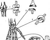
Popular
- Influenza in children: how to treat, what can and cannot be done to parents, what medicines will help?
- "They are people too ..." - you say
- What helps "Prednisolone"
- How are menstruation for endometriosis?
- The fourth stage of cancer, how many live with it
- Prednisolone ampoules instructions for use
- Effective treatments for inflammation of the appendages
- Sinupret - an effective herbal alternative to antibiotics in the treatment of sinusitis
- When do menstruation begin for the first time
- Oncology of the lungs: symptoms and treatment