Pelvic girdle function. Pelvic bones and free lower limb bones
The skeleton of the lower limbs form the bones. pelvic girdle and free lower limb. The pelvic girdle is formed by two pelvic bones. The pelvic bones connected by a sacrum form a pelvis.
\u003e Bones of the pelvic girdle
The pelvis is the basis of the human skeleton. The pelvis is in the middle of the human body. Bottom bones of lower limbs are attached to it, and on top of it rests spinecarrying the head, chest and shoulder girdle with upper limbs. The pelvic bones carry and carry the weight of the entire upper part onto the legs and serve as an attachment point for many powerful muscles of the trunk and legs. The pelvis is formed by two pelvic bones and a sacrum. The pelvic bone consists of three bones: ileal (limit the shape of the pelvis laterally); pubic (limit the shape of the pelvis in front); sciatic
At the junction of these bones is the articular cavity of the hip joint, which includes the head of the thigh. Five intergrown vertebrae of the lower section of the spinal column, forming the sacrum, are located at the bottom and serve as a support for the human body when sitting. All the bones of the pelvis together form a ring, closed behind the sacrum. General form the pelvis resembles an ordinary pelvis, having a cutout in front of the pubic bones. The pelvic axis is tilted forward. The main characteristic points important for determining the proportions and the spatial position of the pelvis are the pubic joint, the protrusions of the crests of the iliac bones and the lower vertebra of the coccyx. When focusing on one leg, the pelvic centerline and the axial line of the shoulders are at an angle to each other.
\u003e Bones of the free lower limb
The bones of the free lower limb are represented by the femur, the two bones of the leg, and the bones of the foot.
Femur. It is the longest tubular bone of the skeleton, it has a body and 2 ends. The upper end consists of a head, a long neck and knolls. The bone is jointed with the pelvis. Behind the neck of the thigh, behind, is a large skewer, which serves as a place of attachment of the muscles of the buttocks. On the inside is a small skewer.
Tibia. At the upper end has concave grooves - the articular surfaces that correspond to the femoral condyles. The lower end of the tibia has an internal ankle, clearly visible on the human body.
Fibula. Its upper edge adjoins the tibia under its external condyle. It is thinner than the tibia, in the upper part ends with the head, and in the lower part - with the external ankle.
Bones of the foot. The foot consists of three main parts: the tarsus, the metatarsus and the phalanges of the fingers. Tarsus consists of seven bones: the ramus, heel, scaphoid, three sphenoid, cuboid. The plyus consists of five short bones lying in one row. Phalanxes of fingers. On the thumb - two phalanxes, on the rest - three each.
Everyone can perfectly imagine the human skeleton, thanks to the numerous photographs and drawings that each of us saw in school. But do we know that the skeleton of an adult person consists of a large number of different boneseach of which performs a specific function?
The human skeleton: what is it made of?
The skeleton of a person is his support. He is not only able to act for the human body as a repository for his internal organs and systems, but also is the place of attachment of its muscles. With the help of a skeleton, a person is able to perform various movements: walk, jump, sit, lie down and much more. An interesting fact is that the human skeleton - the connection of bones - is formed in a child who is still in the womb. True, at first it is only replaced in the process of his life on the bone. In a baby, the bones have practically no hollow space inside. It occurs there in the process of human growth. One of the most important functions of the human skeleton is the formation of new blood cells, which are produced by the bone marrow, which is located in it. The peculiarity of the bones of the human skeleton is the preservation of a certain form during life (and therefore continuous growth and development). The list of human skeletal bones includes more than 200 items. Many of them are doubles, the rest does not form pairs (33-34 pieces). These are some of the bones of the sternum and skull, as well as the tailbone, the sacrum, the vertebrae.
Human limb functions
It is very important to know that the process of evolution, that is, the continuous development of man, left a direct imprint on the functioning of many of his bones. 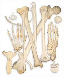 The upper part of the human skeleton with its moving limbs is intended mainly for human survival in the world. With his hands, he is able to cook, do homework, serve himself, etc. Also, there are bones of the human lower limbs. Their anatomy is so well thought out that a person is able to be held upright. At the same time, they serve as the basis for movement and support. It should be noted that the lower limbs are less mobile compared to the upper ones. They are by weight and density more massive. But along with this, their functions are very important for a person.
Human skeleton
Consider the human skeleton: the skeleton of the lower limb and upper limb represented by a belt and a loose part. In the upper section are the following bones: the chest belt, shoulder blades and collarbone, brachial bone and forearm bones, brush. Human bones of the lower limb include: the pelvic girdle (or paired pelvic bones), the thigh, the shin, and the foot. The bones of the free lower limb of a person, as well as the belts, are capable of supporting the weight of a person, therefore, they are so important for him. After all, in fact, only with the help of these connections can it be in a vertical position.
Pelvic girdle (paired pelvic bones)
The first component, which is the basis, forming the bones of the belt of the human lower limb, will be the pelvic bone.  It is she who changes her structure after puberty of any adult. Until this age, it is said that the pelvic girdle consists of three separate pubic and sciatic), interconnected by cartilage tissue. Thus, they form a kind of depression where the head of the thigh is placed. The bone pelvis is formed by joining the anterior bones of the same name. Behind it is articulated by means of the sacrum. As a result, the pelvic bones form a kind of ring, which is a repository for human internal organs.
Femur and Patella
The bones of the belt of the lower limb of a person are not as mobile as the rest of it, which is so called, the free lower limb. It consists of: thigh, shin and foot. The thigh, or femur, is a tubular bone. It is also the largest and longest of all the bones with which the human body is endowed. In its upper part, the femur connects to the pelvic girdle with the help of the head and a long thin neck. Where the cervix passes into the main part of the femur, there are two large bumps on it. It is here that the bulk of the muscles of the human lower limbs is attached. Down the femur becomes thicker. Here are two elevations, through which the thigh is connected, as a result, with the patella and the shin. The patella is a flat bone of a rounded shape, with which the knee is bent. The bones of the lower limb of a person, namely the thigh and patella, carry the following functions: the place of attachment of the bulk of the muscles located on the legs, and the possibility of bending the leg.
Shin
It consists of two bones: tibial and peroneal. They are located next to each other.  The first of them is quite massive and thick. From above, it connects to the outgrowths (condyles) of the femur and the head of the fibula. To the bottom, the tibial bone is transformed on one side into the medial ankle, and on the other, it is located directly under the skin. The fibula is smaller in size. But at the edges it is also thickened. Due to this, it connects from above to the tibia, and from below forms the lateral ankle. It is important that both components of the lower leg, which are also the bones of the human lower limb, are tubular bones.
Human foot bones
The bones are divided into three main parts: the bones of the tarsus, metatarsus and phalanges of the fingers. It is important to note that the foot represents loose bones human lower limb. The first of these include seven bones, the main ones being the bone, called the ram and forming and heel bone. The bones of the metatarsus are further located. There are only five of them, the first one is much thicker and shorter than the others. The toes of the foot are made up of bones called phalanges. The peculiarity of their structure is that the big toe contains 2 phalanges, the other fingers - three pieces each.
Anatomy of the joints of the human lower limbs. Sacroiliac joint, pubic symphysis
Immediately I want to say that all the joints of the lower limb are very large, compared with the joints of the upper limbs. 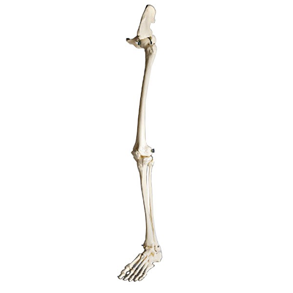 They have a large number of different ligaments, due to which the variety of movements that can be done with the help of human legs. The bones and joints of the bones of the lower extremity were originally created in order to support and move the human body. Therefore, of course, they are reliable, strong and able to withstand heavy loads. Let's start with the top, by location, joints. With their help, the pelvic bones are connected, and a pelvis is formed in humans. In front, such a joint is called the pubic symphysis, and behind it is the sacroiliac. The first one is based on the direction towards each other. Strengthening pubic symphysis formed by a large number of bundles. The sacroiliac joint is very strong and practically immobile. It is tightly fastened not only with the pelvic bones, but also with the lower spine with the help of dense ligaments.
Human pelvis: large and small. Hip joint
It has already been described above that the bones of the girdle of the lower limb of a person are represented primarily by the pelvic bones. They, connecting with the sacrum and pubic symphysis, form a pelvis. This, figuratively speaking, is a ring that protects all organs, vessels and nerve endings inside from external influences. Distinguish large and small pelvis. In women, it is much wider and lower than in men. In the fair sex, everything is thought out to facilitate the generic process, so the pelvis has a more rounded shape and greater capacity. 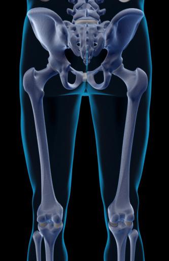 The joints of the lower limb bones are also represented by one of the most well-known representatives of this group, the hip joint. Why is he so famous? Dislocation of the hip joint is the most well-known defect in the development of the lower extremities, which can be identified literally one month after the birth of a baby. It is very important to do it in time, as this untreated diagnosis can bring a lot of trouble in adulthood. The hip joint consists of the cavity of the pelvic bone and the head of the femur. The investigated joint has many ligaments, thanks to which it is strong and quite mobile. Usually experienced orthopedists can diagnose an abnormal development of the hip joint in children through routine examination of the patient. Leaning the legs to the side in a supine position at 180 degrees is possible only with healthy hip joints.
Knee-joint
Imagine a human skeleton. The connection of bones in the form of joints is necessary for a person for the strength of the connection of bones and the creation of maximum mobility of all his limbs. An excellent example of such a connection is He, by the way, is considered the largest joint in the human body. And its structure is very complex: the knee joint is formed with the help of condyles, patella, tibial condyles. The entire joint is shrouded in reliable ligaments, which, along with ensuring the movement of the leg, keep it in the desired position. Thanks to him, not only standing, but also walking. The knee joint can produce various movements: circular, flexor and extensor.
Ankle joint
This joint serves to directly connect the foot and lower leg. Around there are numerous ligaments that provide a variety of movements and the necessary stability of the human body.
Plusphalangeal joints
Studied joints are interesting in their form, compared with other joints of the lower limb of a person. They look like a ball. Strengthening for them are bundles on the sides and on the sole of the foot. They can move, although the diversity of their movement does not differ: small leads to the sides, flexion and extension. Human foot consists of numerous (sedentary) joints and ligaments. With their help, and the movement is carried out, while the human body has the necessary support. So, we can conclude that the bones of the belt of the lower limb of a person are less mobile than the free bones of the same section. But the functions of this no less than those of none.
How do limbs develop with age?
We all know that the human skeleton undergoes certain transformations during life. The skeleton of the lower limb undergoes strong changes with age. Bones that develop on the basis of connective tissue have three stages of their change: connective tissue, cartilage and bone tissue.  Pelvic bone: it is laid in the intrauterine fetal development. Formed cartilaginous layers between the pelvic bones are usually preserved until puberty of a person. Next, they stiffen. Patella: ossification points can appear in a child by the age of 2, completely this happens at about 7 years. Interestingly, the lower limbs in newborns grow much faster than in adults. The peak of such rapid growth falls on the period of puberty: for girls - 13-14 years; boys - 12-13 years.
Remember that the human skeleton is subject to various injuries in the form of damage and even fractures. Since he is entrusted with the implementation of such a large number of important functions of the body, it must be protected. Eating right (food with sufficient calcium content helps strengthen the skeleton), lead an active lifestyle (physical education and sports), monitor your health (any disruption in the functioning of the skeleton to check with a competent specialist) - all this needs to be done by everyone. And then you will meet your old age vigorous, healthy and cheerful.
The pelvic girdle, or the girdle of the lower limbs (cingulum membri inferioris) of a person is formed on each side of a single pelvic bone (os coxae), which, in turn, is the accretion of three bones - ileal, sciatic and pubic. These three bones grow together in the area acetabulum (acetabulum), while the ilium forms the upper side of the cavity, the pubic - lower and anterior, sciatic - lower and back. The acetabulum is circumferentially circumscribed by a protrusion, which, however, in one place (on the medial side) has a notch - this acetabulum tenderloin (incisura acetabuli). The acetabulum is designed to articulate with the femur; for this it has semilunar surface (facies lunata); in the center of the depression is located acetabulum fossa (fossa acetabuli).

Ilium (os ilium)
In the ilium, there are two parts - the lower and upper. The lower part is involved in the formation of the acetabulum and is thickened - this ileal body (corpus ossis ilii). The top one looks like a wide plate - this is iliac wing (ala ossis ilii). In the center the wing is thinner, at the periphery it is thickened; this thickening is called iliac crest (crista iliaca) and has three rough lines for attaching the abdominal muscles - these lines are called outer lip (labium externum), intermediate line (linea intermedia) and inner lip (labium internum). Moreover, the iliac crest has several protrusions - the iliac spines. Is located in front superior anterior iliac spine (spina iliaca anterior superior) and lower anterior iliac spine (spina iliaca anterior inferior). Rear protrusions are called, by analogy, upper posterior iliac spine (spina iliaca posterior superior) and lower posterior iliac spine (spina iliaca posterior inferior).
As for the central part of the wing of the Ilium, it has two surfaces, one external and one internal. The outer surface bears three rough lines - this front, the back and lower gluteal lines (linea glutea anterior, linea glutea posterior and linea glutea inferior); These lines are designed for gluteus muscles. The inner surface has a recess - iliac fossa (fossa iliaca), which is limited from below arched line (linea arcuata). Behind this line is located ear surface (facies auricularis), articulating with the sacral vertebrae; above her - iliac tuberosity (tuberositas iliaca).
The pubis (os pubis)
The pubis is formed body pubic bone (corpus ossis pubic) involved in the formation of the acetabulum coming from it upper branch of the pubic bone (ramus superior ossis pubic), fused with the iliac bone to form iliac-pubic eminence (eminentia illopubica), and lower branch of the pubic bone (ramus inferior ossis pubic), which is the front, downward curved part of the upper branch. The medial bone edge forms symphysial surface (facies symphysialis), connecting with the same surface of the opposite pubic bone - this is how the pubic symphysis is formed. The upper branch also carries the medial end pubic tubercle (tuberculum pubicum), and the lower branch on its back surface is retainer furrow (sulcus obturatorius).
Ischium (os ischii)
Ischium formed sciatic bone (corpus ossis ischii) involved in the formation of the acetabulum, as well as branch of the ischium (ramus ossis ischii), located anterior to the body at an angle, the apex of which is directed backwards. In the area of the specified vertex there is sciatic hill (tuber ischiadicum), and above it - ischial spine (spina ischiadica). Between the sciatic tubercle and the sciatic spine, thus, is formed small sciatic notch (incisura ischiadica minor), and on top of the ischial spine - large sciatic notch (incisura ischiadica major).
The branch of the sciatic bone, moreover, is connected to the lower branch of the pubic bone; thus, between these two branches, as well as the bodies of both of these bones, is formed retainer hole (foramen obturatum).
Skeleton of the lower limbs consists of pelvic girdle and skeleton free lower limbs (legs). The pelvic girdle on each side is formed by an extensive pelvic bone.
Skeleton belt of the lower limbs form two pelvic bones and a sacrum with the tailbone. TO free lower limb bonesinclude: femoral, bones of the leg and foot. The bones of the foot, in turn, are subdivided into the bones of the tarsus, metatarsus and phalanges of the fingers.
Skeleton of the lower limb, right. A - front view; B - rear view; 1 - pelvic bone (os coxae); 2 - femur (femur); 3 - patella (patella); 4 - tibia (tibia); 5 - fibula; 6 - foot bones (ossa pedis)
Pelvic bone (os coxae) in children consists of three bones: ileal, pubic and sciatic, connected in the area of the acetabulum by cartilage. After 16 years, the cartilage is replaced by bone tissue and a monolithic pelvic bone is formed.
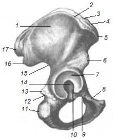
Pelvic bone, right; inside view. 1 - upper posterior iliac spine (spina iliaca posterior superior); 2 - lower posterior iliac spine (spina iliaca posterior inferior); 3 - auricular surface (facies auricularis); 4 - arcuate line (linea arcuata); 5 - large sciatic notch (incisure ischiadica major); 6 - the body of the sciatic bone (corpus ossis ischii); 7 - sciatic spine (spina ischiadica); 8 - small sciatic notch (incisura ischiadica minor); 9 - locking hole (foramen obturatum); 10 - sciatic tuber (tuber ischiadicum); 11 - the branch of the sciatic bone (ramus ossis ischii); 12 - lower branch of the pubic bone (ramus inferior ossis pubis); 13 - symphysial surface (facies symphysialis); 14 - upper branch of the pubic bone (ramus superior ossis pubis); 15 - pubic crest (crista pubica); 16 - body of the pubic bone (corpus ossis pubis); 17 - the body of the ileum (corpus ossis ilii); 18 - the lower front iliac spine (spina iliaca anterior inferior); 19 - superior anterior iliac spine (spina iliaca anterior superior); 20 - iliac fossa (fossa iliaca); 21 - iliac tuberosity (tuberositas iliaca)
Pelvic bone, right; outside view. 1 - iliac crest (crista iliaca); 2 - superior anterior iliac spine (spina iliaca anterior superior); 3 - lower anterior iliac spine (spina iliaca anterior inferior); 4 - acetabulum (acetabulum); 5 - cut of the acetabulum (incisura acetabuli); 6 - pubic tubercle (tuberculum pubicum); 7 - locking hole (foramen obturatum); 8 - sciatic tuber (tuber ischiadicum); 9 - small sciatic notch (incisura ischiadica minor); 10 - sciatic spine (spina ischiadica); 11 - large sciatic notch (incisura ischiadica major); 12 - lower posterior iliac spine (spina iliaca posterior inferior); 13 - lower gluteal line (linea glutea inferior); 14 - upper posterior iliac spine (spina iliaca posterior superior); 15 - anterior gluteal line (linea glutea anterior); 16 - posterior gluteal line (linea glutea posterior)
Ilium (os ilium) - the largest part of the pelvic bone, is its upper section. It distinguishes a thickened part - the body and the flat section - the wing of the Ilium, ending with a crest. On the wing, there are two protrusions on the front and back: the upper anterior and lower anterior iliac spines are in front, and the upper anterior and lower posterior iliac spines are behind. The superior anterior iliac spine is palpable. On the inner surface of the wing there is an iliac fossa, and on the gluteal (outer) - three rough gluteal lines - the anterior posterior and inferior. From these lines the gluteal muscles begin. The back of the wing is thickened, on it there is an ear-shaped (articular) surface for articulation with the sacrum.
Pubic bone (os pubis) is the front of the pelvic bone. It consists of a body and two branches: upper and lower. On the upper branch of the pubic bone is the pubic tubercle and the pubic crest, which passes into the arcuate line of the Ilium. At the junction of the pubic bone with the ileum there is an iliac-pubic eminence.
Ischium (os ischii) forms the lower part of the pelvic bone. It consists of a body and a branch. The lower part of the bone branch has a thickening - sciatic tubercle. At the rear edge of the bone body is a protrusion - the sciatic spine, separating the large and small sciatic notches.
The branches of the pubic and sciatic bones form a obturator opening. It is closed by a thin connective tissue locking membrane. In its upper part there is a obturator canal bounded by a obturator groove of the pubic bone. The channel serves for the passage of vessels of the same name and nerve. On the outer surface of the pelvic bone, at the junction of the bodies of the ileum, pubic and ischial bones, a significant depression is formed - the acetabulum.
Pelvis as whole. The pelvis (pelvis) is formed by the pelvic bones, the sacrum, the coccyx, and their joints.
There are large and small pelvis. The boundary line separating them runs from the cape of the spine along the arcuate lines of the iliac bones, then along the upper branches of the pubic bones and the upper edge of the pubic symphysis. The large pelvis is formed by the deployed wings of the iliac bones and serves as a support for the internal organs of the abdominal cavity. The pelvis is formed by the pelvic surface of the sacrum and coccyx, sciatic and pubic bones. It distinguishes between the upper and lower apertures (entrance and exit) and the cavity. In the pelvis are the bladder, rectum and internal genital organs (uterus, fallopian tubes and ovaries in women; prostate gland, seminal vesicles and spermatic ducts in men).
Genital differences are revealed in the structure of the pelvis: the female pelvis is wide and short, the wings of the iliac bones are greatly expanded. The angle between the lower branches of the pubic bones — the underhead angle — is dull, the cape almost never protrudes into the cavity of the pelvis, the sacrum is wide, short and flat. These features are due to the value of the female pelvis as a generic canal. In the obstetric practice, parameters of the large and small pelvis are used to characterize the pelvis.
Female pelvis; view from above. 1 - boundary line (tinea terminalis); 2 - anatomical conjugate, or straight diameter (diameter recta), of the small pelvis; 3 - transverse diameter (diameter transversa) of the pelvis; 4 - oblique diameter (diameter obliqua) of the pelvis
Female pelvis; bottom view (obstetric position). 1 - direct size of the exit of the small pelvis; 2 - the transverse size of the exit of the small pelvis
The size of the large pelvis of a woman. 1 - ridge distance (distantia cristarum); 2 - spinous distance (distantia spinarum); 3 - spitting distance (distantia trochanterica)
The size of the pelvis of a woman. 1 - true, or obstetric, conjugate (conjugata vera); 2 - external conjugate (conjugata externa); 3 - diagonal conjugate (conjugata diagonal); 4 - direct size of the exit of the small pelvis (diameter recta)
Femur (femur) - the longest bone of the human body. It distinguishes the body, proximal and distal ends. The spherical head at the proximal end faces the medial side. Below the head is the neck; it is located at an obtuse angle to the longitudinal axis of the bone. At the place of transition of the cervix to the bone body there are two protrusions: the large spit and the small spit (trochanter major and trochanter minor). The big spit lies outside and is palpable. Between the spits on the back surface of the bone, the intertroke ridge passes, along the anterior surface, the intertrockle line.
Femur, right. A - rear view; B - front view; B - left view; 1 - femoral head (caput ossis femoris); 2 - femoral neck (collum ossis femoris); 3 - the big spit (trochanter major); 4 - small skewer (trochanter minor); 5 - spitting fossa (fossa trochanterica); 6 - intertrochanic crest (crista intertrochanterica); 7 - gluteal tuberosity (tuberositas glutea); 8 - medial lip (labium mediate) of a rough line; 9 - lateral lip (labium laterale) of a rough line; 10 - fossa musculoskeletal fossa (fossa intercondylaris); 11 - medial condyle (condylus medialis); 12 - lateral condyle (condylus lateralis); 13 - medial epicondyle (epicondylus medialis); 14 - lateral epicondyle (epicondylus lateralis); 15 - the body of the femur (corpus femoris); 16 - rough line (linea aspera); 17 - intertrochanter line (linea intertrochanterica); 18 - fossa of the femoral head (fovea capitis ossis femoris)
The body of the femur is curved, the bulge facing anteriorly. The front surface of the body is smooth, along the back surface there is a rough line. The distal end of the bone is somewhat flattened anteriorly to the rear and ends in the lateral and medial condyles. Above them, respectively, the medial and lateral epicans rise from the sides. Between the latter is located behind the fossa, in the front - the patella surface (for articulation with the patella). Above the inter-latin fossa is a flat, triangular-shaped popliteal surface. The femoral condyles have articular surfaces for joining the tibia.
Patella (patella), or patella, is the largest sesamoid bone; it is enclosed in the quadriceps tendon and is involved in the formation knee joint. It distinguishes the extended upper part - the base and the narrowed, facing down part - the top.
Shin bones: tibial, located medially, and fibular, occupies a lateral position.
Shin bones, right. A - front view; B - rear view; B - right side view; I - tibia (tibia); 1 - upper articular surface (fades articularis superior); 2 - medial condyle (condylus medialis); 3 - lateral condyle (condylus lateralis); 4 - the body of the tibia (corpus tibiae); 5 - tibial tuberosity (tuberositas tibiae); 6 - medial margin (margo medialis); 7 - cutting edge (margo anterior); 8 - intercostal margin (margo interosseus); 9 - medial ankle (malleolus medialis); 10 - lower articular surface (facies articularis inferior). II - fibula: 11 - body of the fibula (corpus fibulae); 12 - head of the fibula (caput fibulae); 13 - cutting edge (margo anterior); 14 - lateral ankle (malleolus lateralis); 15 - inter-muscular elevation (eminentia intercondylaris); 16 - line of soleus muscle (linea m. Solei)
Tibia (tibia) consists of a body and two ends. The proximal end is much thicker; there are two condyle: medial and lateral, articulated with femoral condyles. Between the condoms is an inter-muscular elevation. On the outer side of the lateral condyle is a small fibular articular surface (for connection with the head of the fibula).
The body of the tibia triangular shape. The front edge of the bone protrudes sharply, at the top it goes into tuberosity. At the lower end of the bone from the medial side there is a downward process - the medial ankle. From the bottom, at the distal end of the bone, there is an articular surface for combination with the talus, on the lateral side — a fibular cutting (for joining to the fibula).
Fibula (fibula) - relatively thin, located outwards from the tibia. The upper end of the fibula is thickened and is called the head. On the head there is a tip facing outwards and backwards. The head of the fibula articulates with the tibia. The body of the bone has a triangular shape. The lower end of the bone is thickened, it is called the lateral ankle and is adjacent to the talus bone outside. The edges of the bones of the leg, facing each other, are called interosseous; an interosseous membrane (membrane) of the tibia is attached to them.
Foot bones tarsal bones, metatarsal bones and phalanxes (fingers) are divided into bones.
The bones of the foot, right; back surface. 1 - talus (talus); 2 - talus block (trochlea tali); 3 - head of the talus (caput tali); 4 - calcaneus (calcaneus); 5 - calcaneus tuber (tuber calcanei); 6 - navicular bone (os naviculare); 7 - sphenoid bones (ossa cuneiformia); 8 - cuboid bone (os cuboideum); 9 - metatarsus (metatarsus); 10 - bones of toes (ossa digitorum pedis)
Tarsus bones refer to short spongy bones. There are seven of them: ankle, heel, cuboid, scaphoid, and three wedge-shaped. The talus has a body and a head. On the upper surface of her body is a block; together with the bones of the lower leg, it forms the ankle joint. The calcaneus is located under the talus, the largest of the tarsus bones. On this bone, there is a well-pronounced thickening - the calcaneal tuber, the process called the support of the talus, the ram and cuboid articular surfaces will serve to connect with the corresponding bones).
The cuboid bone is located in front of the calcaneus, and the navicular bone lies anterior to the head of the talus. Three wedge-shaped bones - medial, intermediate and lateral - are located distal to the navicular bone.
Metatarsus in the amount of five are located anterior to the cuboid and wedge-shaped bones. Each metatarsal bone consists of a base, body, and head. By their bases, they are articulated with the bones of the tarsus, and with their heads with proximal phalanges of the fingers.
Toes, like fingers, have three phalangesexcept for the first finger, which has two phalanges.
The skeleton of the foot has features due to its role as part of the support apparatus in the vertical position of the body. The longitudinal axis of the foot is almost at a right angle to the axis of the leg and thigh. In this case, the bones of the foot do not lie in the same plane, but form a transverse and longitudinal arches, turned with a concavity towards the sole, and convexity toward the rear of the foot. Due to this, the foot rests only on the heel of the calcaneus and the heads of the metatarsal bones. The outer edge of the foot below, it almost touches the surface of the support and is called the supporting arch. The inner edge of the foot is raised - this is the spring arch. This structure of the foot ensures that it performs the support and spring functions, which is associated with the vertical position of the body and erect position.
End of work -
This topic belongs to:
The position of man in nature. Anatomy and physiology as a science. Methods for studying the human body
Tissue is a system of cells and intercellular substance having the same structure of origin and function ... Intercellular substance is a product of vital activity of cells. It provides ... Tissue cells have a different shape that determines their function. Tissues are divided into four types ...
If you need additional material on this topic, or you did not find what you were looking for, we recommend using the search in our database:
What we will do with the resulting material:
If this material turned out to be useful for you, you can save it to your page on social networks:
| Tweet |
All topics in this section:
The position of man in nature. Anatomy and physiology as a science. Methods for studying the human body
The position of man in nature. Man is a part of the biosphere, a product of its evolution, therefore the state of his health is closely dependent on the state of the environment. WITH
Parts of the human body. Axes and planes of the human body
Parts of the human body. The structure of the human body is the same as in all mammals. In the human body there are: head, neck, torso and two pairs of limbs. In each part
Anatomical nomenclature. The constitution of man, the morphological types of the constitution. Definition of authority. Organ systems
Anatomical nomenclature. In anatomy adopted Latin terminology, which is used throughout the world. The system of organs, organs and their parts have Latin designations. Aggregate
Muscle tissues: functions, types
Muscle tissue. The motor processes in the human and animal body are due to the contraction of muscle tissue, which has contractile structures. For muscle tissue include n
The internal environment of the body. Homeostasis: its nervous and humoral regulation mechanism. Blood like tissue
Blood like tissue. Blood and lymph are connective tissues with special properties. They are called tissues of the internal environment. Due to the constant blood circulation is provided: 1
The process of hemopoiesis. The functions of the blood - transport and protective. Blood Composition: Plasma and Uniform Elements
Hemopoiesis (blood formation) is the process of formation, development and maturation of blood cells - leukocytes, erythrocytes, and platelets in vertebrates. Organ systems involved in
The study of blood cells. Erythrocytes: structure and function. Erythrocyte norm. Hemoglobin
Uniform elements (40% of blood): · 96% - red blood cells - carry oxygen and carbon dioxide; · 3% - leukocytes - cells of the immune system; ·
Platelets: structure, function, norm. Hemostasis. Blood coagulation factors
Platelets, or blood plates, are colorless spherical bodies devoid of nuclei. Their diameter is 2-3 microns, 3 times smaller than the diameter of red blood cells. Platelets are formed
Rh factor. Designation, localization. The concept of Rhesus conflict. ESR: norms for men and women, diagnostic value
In addition to the main agglutinogens A and B, there may be additional erythrocytes, in particular the so-called Rh factor (Rh-factor), which was first found in the blood of the monkey monkey Rh
The human skeleton: functions, departments
The skeleton is a collection of bones belonging to them of the cartilage and ligaments connecting the bones. There are more than 200 bones in the human body. The weight of the skeleton is 7-10 kg, which is 1/8 of the human weight.
The connection of the bones. Joint structure
The connection of the bones. All the bones in the human body are interconnected in various ways into a coherent system - the skeleton. But all the variety of bone connections in the skeleton can be reduced to two
Divisions of the skull. Brain bones
Divisions of the skull. The skull (cranium) consists of the brain and facial. All bones are interconnected relatively motionless, except lower jawforming a combo
The bones of the facial skull
TO facial skulls (splanchnocranium) include 15 bones: · unpaired - lower jaw, vomer, hyoid bone; · Paired - upper jaw, palatine, with
The skeleton of the body - the structure of its components. Features of the structure of the thoracic, cervical, lumbar vertebrae, sacrum, tailbone. Spinal movements
The human skeleton consists of four sections: the skeleton of the trunk, the skeleton of the head (skull) and the skeleton of the upper and lower extremities. The skeleton of the trunk includes the spinal column and bones.
Spinal column - departments, the number of vertebrae in them. Physiological curves of the spine, their formation, meaning
According to its development, the vertebral column (columna vertebralis) forms around the spinal cord, forming a bone container for it. Apart from protecting the spinal cord, the spinal column is in
The belt of the upper limbs. Skeleton of the upper limbs
The skeleton of the upper limbs consists of shoulder girdle and the skeleton of the free upper limbs (arms). The structure of the shoulder girdle consists of two pairs of bones - the clavicle and scapula. TO
Muscles of the head: chewing and mimic - their features and functions
Chewable muscles. Chewing include the temporal, chewing, medial and lateral pterygoid muscles. They are differentiated from the musculature of the first visceral (maxillary) du
The median group. Subhyoid muscles
1. Double-abdominal muscle has two abdomen - anterior and posterior, connecting with a tendon web. Start: anterior abdomen - double-abdominal fossa of the lower jaw, posterior - mastoid temporal fossa
Subhyoid muscles
1. The scapular-hypoglossal muscle is long, thin, and is divided by an intermediate tendon into two abdomens. Start: upper abdomen - lower edge of the hyoid bone, lower - upper edge of the scapula, ver
Medial group
1. The long neck muscle consists of two parts - the lower (medial) and upper (lateral). Start: lower - the bodies of the three upper thoracic and three lower cervical vertebrae, the upper - the transverse
Neck fascia
The anatomy of the fasciae of the neck due to the large number of organs and muscles in this area of the body is quite complex. Cervical fascia is divided into three plates: superficial, pretracheal and prevertebral.
Muscles of the trunk: chest, back and abdomen
Chest muscles. Own musculature of the thoracic region of the trunk, which lies in the depths, preserves, as the skeleton of this region, a segmental structure. Muscles are arranged in three layers: 1)
Muscles of the upper limb girdle
Chest muscles. Own musculature of the thoracic region of the trunk, which lies in the depths, preserves, as the skeleton of this region, a segmental structure. Muscles are arranged in three layers: 1) external intercostal space.
Muscles of the upper limbs
The muscles of the shoulder girdle. Shoulder girdle, firming free limb on the body, it connects with only one sternoclavicular joint. The strengthening of the shoulder girdle is performed by the muscles.
Muscles of the lower limb girdle
The muscles of the pelvic girdle. The pelvic girdle is almost motionlessly articulated with the sacral spine, therefore there are no muscles that set it in motion. The muscles located on the pelvis lead
Muscles of the lower limbs
Thigh muscles. From the wide fascia on the sides of the thigh, two dense intermuscular septa extend to the femur along its rough line. Together with the third thin fascial
The value of oxygen and carbon dioxide for humans. Breathing process - definition, stages
Breathing is the exchange of gases, on the one hand, between the blood and the external environment (external respiration), on the other - the exchange of gases between the blood and the cells of the tissues (internal or tissue respiration). Mediator
Overview of the respiratory system: airways and lungs, their functions and structure
Respiratory organs include the nasal cavity, the larynx, the trachea, the bronchi and the lungs. In the respiratory system, airways (respiratory) passages (nasal cavity, larynx, trachea and bronchi) and respiratory
Nasal cavity: structure and function
The path along which the inhaled air passes, begins with the nasal cavity. The nose is a complex formation. It consists of an external nose and nasal passage, its walls are formed
Larynx - projection on the spine, the structure and function of the larynx
The larynx is the upper part of the vocal tube, serves to hold air and is a sound-forming organ. It is located in the front of the neck, at the top is suspended from the hyoid bone, is located
Additionally
Throat cartilage. 1. The cricoid cartilage, cartilago cricoidea, hyaline, forms the base of the larynx. It is similar in shape to a ring and consists of a plate, lamina cartilagini
Bronchial tree
The right and left main bronchi begin at the trachea bifurcation at the level of the upper margin V thoracic vertebra and go to the gate of the right and left lungs, respectively. In the area of the gates of the lungs each
Pleura - structure, leaflets, pleural cavity, pleural sinuses, pressure in the pleural cavity
The pleura (pleura) is a very thin, smooth and moist serous membrane rich in elastic fibers, lined on the free surface with mesothelium (single-layer flat epithelium
Oral cavity - threshold and proper oral cavity
The oral cavity (cavitas oris) is subdivided into two sections: the mouth of the mouth and the oral cavity itself (Fig. 54). The eve of the mouth is a slit-like space located between the lips and u
Zev - borders, palatine arches, soft palate. Tonsil Lipoepithelial Ring
Zev (isthmus faucium) is a space bounded by the palatine arches, the soft palate and the root of the tongue. It reports oral cavity with a throat. In the front arch laid m. palatoglossus and in the back - m. p
Oral cavity organs: tongue, teeth
The tongue, lingua (Greek glossa, hence the inflammation of the tongue - glossitis), represents the muscular organ (streaked arbitrary fibers). Changing its shape and position matters to
Pharynx, its topography, wall structure, divisions, functions
Pharynx, Pharynx, represents that part of the digestive tube and respiratory tractwhich is the connecting link between the nasal cavity and the mouth, on the one hand, and the esophagus and the larynx, with each other
Esophagus: location, departments, physiological narrowing, wall structure, function
The esophagus (esophagus) is a narrow, long tube that connects the pharynx to the stomach. It begins on the neck, at the level of the VI cervical vertebra, and ends in the abdominal cavity, at the level of the XI thoracic
Functions of the esophagus
Of course, the main function of the esophagus is transport, or motor. The main task of the esophagus is the speedy delivery of food to the stomach, without mixing it. Move pi
Pancreas - location, structure, function. The composition of the digestive juice. Regulation of pancreatic juice excretion
The pancreas, pancreas, is the second largest digestive gland, as well as the endocrine gland. The pancreas is an elongated organ with
Pancreas Functions
The excretory function of the pancreas is to release pancreatic juice into the duodenum, which contains enzymes (trypsin, lipase, maltose, lactase, etc.), neutralizing
Gallbladder - location, structure, function. The composition and properties of bile, function of bile, types of bile (hepatic, cystic)
The gallbladder, vesica fellea is a reservoir in which bile accumulates. It is located in the fossa of the gallbladder on the visceral surface of the liver, has a pear shape.
The composition of bile
Bile consists of 98% water and 2% of the dry residue, which includes organic substances: bile salts, bile pigments - bilirubin and biliverdin, cholesterol, fatty acids, lecithin, mucin, urea
Suction in the small intestine
Under the suction understand the process of transition of water and dissolved in it nutrients, salts and vitamins from the alimentary canal into the blood and lymph. Absorption mainly occurs in the small intestine.
Heart - the location, structure, projection on the surface of the chest. Heart chambers, heart holes. Valves of the heart - the structure and function.
The heart is a hollow muscular organ having the shape of a cone, 250-360 g, in newborns - 25 g. Located in the chest cavity, behind the sternum, in the anterior mediastinum: 2/3
Heart wall structure
The walls of the cavities of the heart vary in thickness, in the atria 2-5 mm, in the left ventricle approx. 15 mm, in the right approx. 6 mm. 3 layers: internal ENDOCARD (flattened, thin, smooth endothelium) - lined
Cardiac conduction system
Regulation of heart activity depends on the needs of the body and is carried out by the vegetative nervous system. Sensory and motor nerve fibers pass to the heart as part of a wandering
Cardiac cycle - its phases, the duration of the cardiac cycle. External manifestations of the activity of the heart - heart impulse, heart tones
Cardiac cycle. The heart of a healthy person contracts rhythmically, in conditions of rest with a frequency of 60 - 70 per minute. During muscular work, when the body temperature rises or surround
Aorta - divisions, topography, areas of the blood supply. Arteries of the neck and head. Blood supply to the brain
The aorta (aorta) is the largest human arterial vessel, the main line from which all the arteries of the body originate. Departments. Ascending in the aorta
Arteries of the upper limbs
The right subclavian artery departs from the brachial trunk, the left - from the aortic arch. Each artery first goes under the collarbone above the dome of the pleura, then passes into the gap between the front and middle stairs.
Lower limb arteries
The continuation of the aorta in the pelvis is a thin median sacral artery. The right and left common iliac arteries are the terminal branches of the abdominal aorta. At the level of the sacroiliac
The system of the inferior vena cava, the area of outflow of blood in it
The system of the inferior vena cava is formed by vessels collecting blood from the lower extremities, walls and organs of the pelvis and abdominal cavity. The inferior vena cava is formed from the fusion of the left and right
System superior vena cava. Veins of the head and neck. The veins of the upper limb are superficial and deep. Chest veins
All venous blood from the organs of the body flows to the right, venous, half of the heart through the two largest venous trunks: the superior vena cava and the inferior vena cava. Only own veins of the heart fall.
Abdomen veins - parietal, internal, areas of outflow of blood in them. Venous anastomoses
The unpaired vein, being one of the main ducts of the superior vena cava, begins in the abdominal cavity by merging, among others, the following parietal veins: 1) right ascending
Portal vein system
The portal vein has a length of 4–5 cm and a diameter of 15–20 mm. It is formed behind the head of the pancreas by merging the inferior mesenteric, superior mesenteric, and splenic veins. Then she send
Factors affecting blood circulation. Causes of blood flow in arteries, veins, capillaries. Blood pressure. Pulse, its characteristics
The principle of blood movement. The third principle of hydrodynamics, applied to the blood flow, reflects the law of conservation of energy and is expressed in the fact that the energy of a certain volume of flowing fluid
Spinal cord, its location, structure and shell
The spinal cord lies in the spinal canal and is a cord 41-45 cm long (in an adult), somewhat flattened from front to back. Above it goes directly to the head.
Segment - the concept, species, roots of the spinal cord
The transverse segment of the spinal cord, corresponding to a pair of ventral and a pair of dorsal roots, forms a nerve segment, neurotome. The human spinal cord consists of 31 segments. From each segment
Reflex arc
Reflex - the response of the body to irritation from the external or internal environment, carried out with the participation of the central nervous system. All nervous activity consists of reflexes. varying degrees cl
Brain - location, divisions, parts
The brain is located in the cavity of the skull. Ovoid shape due to protruding frontal and occipital poles. The upper lateral surface is convex, dotted with furrows over which
The shell of the brain and interspaces, location, their contents. Ventricles of the brain. Liquor - education, movement, functions
From the earliest period of its development, the formation of the central nervous system is surrounded on all sides by the mesenchyme, which accompanies all the nervous and gliotic elements as they move to the periphery
Bridge and cerebellum: structure and function
The hindbrain includes the bridge and the cerebellum. It develops from the fourth brain bubble. The bridge below is bordered by the medulla oblongata, above it passes into the legs of the brain, laterally
Mid brain: structure and function
The midbrain includes the ventral ventral legs and the lamina of the roof or quadripoxy, which lies dorsally. The cavity of the midbrain is the aqueduct of the brain. Roof plate consists
Intermediate brain - the structure of it forming, the main functions
The intermediate brain is located under the corpus callosum and the vault, growing together on the sides with the hemispheres of the large brain. These include the thalamus (visual cusps), epithalamus (naduhgorn
The final brain: the structure. Right and left hemispheres, their surfaces, lobes
The terminal, or the big brain develops from the anterior cerebral bladder, consists of strongly developed paired parts - the right and left hemispheres of the large brain and the conjunction
Bark of the big hemispheres. Conditioned-reflex activity of the cortex. The role of the cortex in meeting the needs of the organism in human adaptation
The cerebral cortex is a layer of gray matter up to 4 mm thick, covering the surface of the hemispheres and lying deep in the furrows. The cortex is formed by layers of nerve cells and volokol
Projection zones of the cortex. Associative fields and their functions
In each hemisphere, there are three surfaces: upper lateral - convex, respectively, the cranial vault, medial - flat, facing the same surface of the other hemisphere, and lower - wrong
Cranial nerves, their number, name, place of exit and function. Spinal nerves: formation, number, branches of spinal nerves
From the brain leaves 12 pairs of cranial nerves. Each pair of nerves has a number and a name. In contrast to the spinal nerves, which are all mixed and only divided at the periphery
Thoracic spinal nerves - location, branches, types and areas of innervation of the posterior and anterior branches
The thoracic spinal nerves in an amount of 12 pairs are segmented in the intercostal spaces, below the intercostal arteries, with the first going out between the I and II thoracic vertebrae. 1978 Land
Spinal nerves and plexuses
The human spinal cord consists of 31 segments. A pair of spinal nerves departs from each segment by two roots, anterior and posterior. In the region of the intervertebral foramen, in the posterior root,
Vegetative nervous system (sympathetic and parasympathetic parts)
Vegetative nervous systemconsisting of two parts: sympathetic and parasympathetic, innervates the whole organism, all organs and tissues: glands, muscles of blood vessels and internal
Additional Information
Other physiological schools. In addition to school I.P. Pavlova, successfully developing the conditioned-reflex theory in our time, there are a number of other areas. For example, very widely
Teaching I.P. Pavlov on analyzers. Divisions of the sensory system
All irritations that have an effect on the body are perceived by means of sensitive nerve endings - receptors embedded in special sensory organs (eyes, ear), and in all others
Eye - the structure of the eyeball
The organ of vision, the eye, consists of an eyeball and an auxiliary apparatus. From the eyeball comes the optic nerve connecting it to the brain. The eyeball has the shape of a ball
Auxiliary apparatus. The optical system of the eye - the structures related to it. Conditions for clear vision of objects, factors determining them
The auxiliary apparatus of the eye consists of protective devices, the lacrimal and motor apparatus. The protective formations include eyebrows, eyelashes and eyelids. Eyebrows protect your eyes from sweat.
Departments of the ear, their structure
The pre-cochlear organ, or organ of hearing and balance, is one of the complex sensory organs, since it contains several types of receptor cells: a) receptors that perceive sound vibrations
Olfactory organ. Olfactory receptors. Conductor and central parts of the olfactory sensory system
At the end of the 1st month of development in the ectoderm of the head end of the body, two thickened nasal plates appear, lining the bottom of the nasal fossae. The pits mutually approach each other, forming the nasal cavity. Layer n
The organ of taste. Taste buds. Conductor department. Taste centers
The development of taste bulbs. At the end of the 2nd month of embryo development, cell bundles related to the embryonic nervous glia appear on the tongue. In the 4th month nerve fibers sprout to these cells.
Skin, its structure. Skin glands, skin derivatives: hair, nails
The body of touch is the skin. The skin forms the outer cover of the body, whose area in an adult reaches 1.5-2.0 square meters. The skin is a huge receptor field, in to


Popular
- Breast cancer is curable at any stage.
- The remedy for the cold Sinupret
- Azitrox - official instructions for use
- Chicken-bjaka: allowed antibiotics were found in Russian chicken
- Oral Cancer: Symptoms and Treatment
- Dark and thick blood during menstruation.
- Modern analogues of doxycycline tablets
- Is it possible to die from pneumonia
- What earwax will tell all about your health
- Tussin: instructions for use