What is the nasal cavity? Nasal cavity: functions, structure, sinuses
text_fields
text_fields
arrow_upward
The nasal cavity is the initial part of the respiratory system. In addition, there are olfactory receptors.
The nasal cavity is divided by a septum into two almost symmetrical parts and communicates with the external environment to the nostrils. Their lateral walls are called nose wings and formed by small cartilage.
On top of the nostrils are limited to a pair of large cartilages of the wings of the nose. The base of the external nose, its back, is formed in the area of the root of the nose by the nasal bones and paired lateral cartilage (Fig. 4.28).
Fig. 4.28.
The latter rest in front of the cartilaginous nasal septum (continuation of the bone, formed by the perpendicular plate, ethmoid bone and vomer), and behind - at the nasal bones and edges of the pear-shaped hole formed by the maxillary bones.
The cartilaginous skeleton of the nose keeps the nostrils constantly open, through which the upper airways communicate with the external environment. The protruding nose with the nostrils facing down is a specific feature of the human face.
The nasal cavity is divided by a longitudinal partition on the right and left parts. They communicate with the nasopharynx through the choans (see Atl.). Each half, in turn, is divided into the upper, middle, and lower nasal conchs into the passages into which the pneumatic sinuses of the skull open. In the lower nasal passage, the nasolacrimal canal opens, in the middle - the maxillary (maxillary) and frontal sinuses and front cells of the ethmoid bone, and in the upper - its posterior cells and sphenoid sinuses (see Athl.).
Fig. 4.28. Nose cartilage:
A - from the side;
B - from the bottom;
1 - frontal and 2 - nasal bones;
3 - the frontal process of the upper jawbone;
4 - lateral cartilage;
5 - large cartilages of the nose wings;
6 - small cartilage cartilage;
7 - nostril;
8 - zygomatic bone;
9 - the top of the nose,
10 - the maxillary bone;
11 - nasal septum cartilage.
The mucous membrane of the nasal cavity
text_fields
text_fields
arrow_upward
The mucous membrane of the nasal cavity is firmly adhered to the bone base.and through the corresponding holes penetrates into the sinuses of the bones of the skull. In the posterior regions through the choans, it gradually passes into the mucosa of the nasopharynx and soft palate.
In the mucosa emit:
- respiratory and
- olfactory area.
In the latter are located sensitive endings of olfactory neurons.
Olfactory region occupies the top nasal concha and adjacent parts of the middle shell and septum of the nose. This part of the cavity is phylogenetically older than the respiratory part, since it is already present in vertebrates with gill breathing, which do not have a nasal cavity.
The rest of the nasal mucosa refers to respiratory part. It is richly supplied blood vessels, for the most part is covered with a multi-row ciliated epithelium. In the epithelial cover there are many goblet cells secreting mucus, which, together with dust particles settled on the membrane, is removed by ciliary movements of the cilia. Flowing into the nasal cavity along the lacrimal-nasal duct, the secret of the lacrimal glands increases the hydration of its mucous membrane.
Own mucous plate contains numerous mucous and serous glands. There are also lymphatic vessels and follicles, the most numerous at the entrance to the nasopharynx. In the middle and lower shell in its own plate there is a large number of thin-walled venous vessels, which in normal conditions are in a collapsed state. However, they are able to stretch and overflow with blood. In this case, there is a feeling of nasal congestion.
In the nasal cavity, the inhaled air is heated (or, on the contrary, cooled, if it is very hot), partially cleaned of mechanical impurities (dust, smoke) and moistened. Therefore, it is very important that breathing occurs through the nose, and not through the mouth, as is the case with pathological growths of the nasal mucosa (for example, with polyps), when nasal breathing is difficult.
Cavum nasi, is a space that lies in the sagittal direction from the pear-shaped aperture to the hoan and is divided by a septum into two halves. The nasal cavity is limited to five walls: upper, lower, lateral and medial.
Upper wall formed by frontal bone, the inner surface of the nasal bones, the lamina cribrosa of the ethmoid bone and the body of the sphenoid bone.
Bottom wall formed by the bony palate, palatinum osseum, which consists of the palatine process of the upper jaw and the horizontal plate of the palatine bone.
Lateral wall formed by the body of the upper jaw, the nasal bone, the frontal process of the upper jaw, the lacrimal bone, the labyrinth of the ethmoid bone, the lower nasal shell, the perpendicular plate of the palatine bone and the medial plate of the pterygoid process.
Medial wall, or nasal septum, septum nasi osseum, divides the nasal cavity into two halves. It is formed by the perpendicular plate of the ethmoid bone and the ploughshare, on the top - the nasal spine frontal bone, spina nasalis, behind - wedge-shaped crest, crista sphenoidalis, sphenoid bone, from below - nasal crest, crista nasales, upper jaw and palatine bone. The nasal cavity opens in front of the pear-shaped aperture, apertura piriformis, and behind - with hoans. Choanae, choanae - paired internal openings of the nasal cavity, which connect it to the nasal part of the pharynx.
On the lateral wall of the nasal cavity are three turbinates: the upper, middle and lower, concha nasalis superior, media et inferior. The upper and middle turbinates belong to the labyrinth of the ethmoid bone, the lower is an independent bone. The listed shells limit three nasal passages: upper, middle and lower, meatus nasalis superior, medius et inferior.
Upper nasal passage, meatus nasalis superior, lies between the upper and middle turbinates. It opens the rear cells of the ethmoid bone. At the posterior end of the superior nasal concha, there is a clinopathic foramen, foramen sphenopalatinum leading to the fossa pterygopalatina, and above the superior nasal concha is a wedge-shaped recess, recessus spheno-ethmoidalis, in the region of which the sphenoid of the sphenoid bone, sinus sphenoidalis, opens.
Middle nasal passage, meatus nasalis medius, located between the middle and lower turbinates. Within its limits, after removing the middle shell, the lunate hole opens, hiatus semilunaris. The lower part of the semilunar orifice expands, at the bottom of which there is a hole, hiatus maxillaris, leading to maxillary sinus, sinus maxillaris. In the anterior-upper part of the nasal cavity, the lunate foramen expands and forms a ethmoid funnel, infundibulum ethmoidale, into which the frontal sinus opens, sinus frontalis. In addition, in the middle of the nasal passage and the lunar foramen, the anterior and some middle lattice cells open.
Lower bow, meatus nasalis inferior, located between the bone sky and the lower turbinate. It opens nasolacrimal canalis canalis nasolacrimal. In clinical (otolaryngological) practice, puncture is performed through the lower nasal passage. maxillary sinus for diagnostic and therapeutic purposes.
The slit-like space between the posterior portions of the concha and the bony nasal septum is called the common nasal passage, meatus nasi communis. The section of the nasal cavity, located behind the nasal conchae and the osseous nasal septum, forms the nasopharyngeal passage, meatus nasopharyngeus, which opens into the posterior nasal openings, the choans.
Buttresses - these are bone thickenings in separate parts of the skull, combined with each other by transverse interchanges, through which during chewing the force of pressure is transmitted to the arch of the skull. The buttresses balance the force of pressure that occurs during chewing, jolts and jumps. Between these bulges are located thin bone formations called weak spots. It is here that fractures occur more often during physical activity, which does not coincide with the physiological acts of chewing, swallowing and speech. AT clinical practice fractures in the neck area are more common lower jaw, angle and upper jaw, as well as the zygomatic bone and its arc. The presence of holes, cracks and weak points in the bones of the skull determine the direction of these fractures, which is important to take into account in oral surgery. In the upper jaw, the following buttresses are distinguished: fronto-nasal, collar-zygomatic, palatine, and pterygoidal; at the bottom - cellular and ascending.
In fact, this organ is paired, that is, there are two nasal cavities. They are separated from each other by a nasal septum. Each is opened in front of the nostrils, and behind it is connected with the nasopharynx by specific openings. However, it turned out that these two departments are united in speech under the name " nasal cavity».
Its structure is more complicated than it seems to an unknowing person. The walls of the nasal cavities, the bottom and the roof of the cavity are hard thanks to the bone, cartilage and connective tissue of high density. It is because of this feature of the structure when inhaling the cavity does not collapse.
Each nasal cavity is divided into two parts: the vestibule is an enlarged area immediately behind the nostrils, the respiratory cavity is a narrowed part located immediately after the vestibule. The epidermis, which lines the cavity from the inside, contains a lot of hair follicles, and also sweat and sebaceous glands. What exactly is the nasal cavity lined for? Its functions are cleansing, raising the humidity and temperature of the air, which is why it is also so abundantly humiliated with blood vessels. Hairs can trap large particles in the air we breathe.
In anticipation of the multilayer refers to the non-threshold type, then it becomes a multi-row cylindrical ciliate, in it goblet cells begin to appear. The epithelium becomes part of the mucous lining the respiratory part of the nasal cavity.
Own mucous plate here is adjacent to the periosteum or perchondrium, depending on whether this mucous bone or cartilage covers. The basement membrane, which separates the respiratory epithelium from its own plate, is much thicker than in most other types of epithelium.
The epithelial surface is moistened with mucus, which is produced by the glands from its own mucous plate. During the day up to 500 ml of mucus is produced. The latter is mixed with particles of dirt and dust that stick to it, and thanks to the cilia moves to Cleansing the nasal cavity depends largely on the state of the cilia, if they are affected by illness or injury, this process can be greatly disturbed.
In some places near the vestibule there are lymphatic follicles that perform an immune function. In the lamina propria of the nasal mucosa there are many plasma cells and lymphocytes, and sometimes there are granular leukocytes. They "protect the boundaries" of the body, protecting us from invasions, because the nasal cavity often becomes the gateway of infections.
However, the cavity “works” not only with air, on the upper part of the walls, as well as the roof of the back of each area, there are special cells that make up the organ of smell.
There are two olfactory zones, one in each nasal cavity. The mucosa there forms a special organ, thanks to which we are able to smell. The peculiarity of this sensory organ is that the bodies of neurons there are located on the surface, which makes them really vulnerable. Therefore, with injuries of the nose or chronic diseases a person may lose his sense of smell. We also lose about one percent of our sense of smell each year, which is why this important feeling is so often disturbed in the elderly.
Along the lateral plate of each cavity there are three bone plates that are one above one, like small shelves. They are slightly curved downwards, therefore, they were called the bow conchs.
Sinuses (sinuses), which are located in the bone cavities, are also connected with the nasal cavity. The largest is in the smaller sinuses - in the frontal, ethmoid and sphenoid bones. They are filled with mucus and sometimes with pus in sinusitis. In this case, drugs are prescribed that make the sinuses and patency increases.
The nasal cavity is difficult, because it must protect us, prepare the air for the lungs and carry out the smell.
There are external nose and nasal cavity.
The internal structure of the nose consists of a hard bone part and a soft cartilage part. The nasal bones are located at the top of the nose and are shaped like a pyramid. They form the base of the nose and make up the upper third of the nose. The lower two thirds of the nose is made of cartilage. The cartilage forms the shape of the lower part of the back of the nose and the shape of the tip of the nose. There are two connected cartilaginous structures: the upper lateral cartilage and the lower lateral cartilage (pterygoid cartilage). The upper lateral cartilage connects the nasal bone with the lower lateral cartilage. The lower lateral cartilage is shaped as a curved "C" and has three areas: the outer part (lateral leg), middle part (dome), and the inner part (middle leg). It forms the wings of the nose.
The two middle legs form a jumper between the nostrils, which is called columella.
External nose It looks like a pyramid and is formed by bones, cartilage, muscles. Outside the nose is covered with the same skin as the face. It distinguishes: root, back, apex and wings of the nose. The root of the nose is located in the upper part of the face and is separated from the forehead by the nose. The sides of the nose in the midline are connected and form the back of the nose. Down the back of the nose goes to the top of the nose, below the wings of the nose limit the nostrils leading into the nasal cavity.
The external nose is an important part of the cosmetic face ensemble. In the nasal cavity distinguish the threshold of the nose and its own nasal cavity.
Nasal vestibule covered inside the skin of the external nose, which continues here through the nostrils, the skin of the vestibule contains hairs, sweat and sebaceous glands.
The vestibule passes into the nasal cavity, which is a channel that runs longitudinally through the bones of the facial skeleton and has the shape of a prism. The bottom of the nasal cavity is a hard palate. The nasal cavity is lined with mucous membrane.
Nasal cavity the septum is divided into two halves: the right and left, in the partition there are bone and cartilage parts. Behind the choanal cavity, the nasal cavity communicates with the nasal pharynx. Most of the nasal cavity is represented by the nasal passages with which the paranasal sinuses (the air-cavity of the skull bones) communicate. Three turbinates (upper, middle and lower), located on the side walls, increase the total surface of the nasal cavity. Between the inward-facing surfaces of the apertures and the nasal septum there is a slit-like common nasal passage, and under the shells there are nasal passages having the appropriate names: upper middle and lower. In the lower nasal passage, the nasal duct opens, in the upper - the posterior cells of the ethmoid bone and the sphenoid sinus, in the middle - the middle and anterior cells of the ethmoid bone, the frontal and maxillary sinuses.
Nasal mucosa, it is possible to distinguish two parts differing from each other in structure and function: the respiratory and olfactory. The respiratory part occupies the area from the bottom of the nasal cavity to the middle of the middle turbinate. The mucous membrane of this area is covered with ciliated epithelium and contains a large number of glands secreting mucus, in addition, there are many blood vessels in the submucosa.
The olfactory region is occupied by a part of the nasal mucosa, covering the right and left upper nasal conchs, as well as part of the middle shells and the corresponding division of the nasal septum. In the olfactory region are nerve cells that perceive odorous substances from the inhaled air.
To the paranasal sinuses carry the air-cavity, surrounding the nasal cavity and connected with her holes (excretory ducts). There are maxillary (maxillary), frontal, sphenoid and ethmoid sinuses. Their sizes are different for different people, the maximal sinus is considered to be the largest (from 5 to 30 cm3). Inside the sinuses are also lined with mucous membranes.
The maxillary sinuses are located in the body of the upper jaw, to the right and left of the nasal cavity. The roots of the teeth of the upper jaw (3-6) in some cases may act in the sinus, so it may develop odontogenic inflammatory processes. The frontal sinuses are located in the frontal bone at the level eyebrows right and left. The ethmoid sinuses consist of individual cells and are located in the thickness of the ethmoid bone. The sphenoid sinus is located in the body of the sphenoid bone (behind the ethmoid bone) and is divided by a septum into two halves. Through special holes, the sinus communicates with the nasal cavity.
The nose performs various functions: respiratory, protective, resonator and olfactory.
Respiratory function is the main. The nose first perceives the inhaled air, which is warmed, cleaned and moistened here, so nasal breathing is most physiological for the body.
The protective function is that the receptors of the mucous membrane react to many stimuli from the external environment: chemical composition, temperature, humidity, dust and other air properties. When exposed to the mucous membrane of stimuli, sneezing and tearing appear. A tear that enters the nasal cavity through the nasolacrimal canal enhances the secretion of mucous glands and removes irritating substances from the nasal cavity.
In the mechanical removal of substances suspended in inhaled air, the ciliary epithelium of the nasal mucosa plays an important role. When the cilia oscillate, directed from the entrance to the nose to the nasopharynx, there is a movement of particles that have fallen into the nasal cavity. Some of the larger dust particles are retained in the nasal vestibule, and if the dust particles suspended in the air do get into the nasal cavity, they are removed from it with mucus when sneezing or blowing your nose. The protective mechanisms also include warming and moistening the incoming air through the nose.
The resonator function is provided by the presence of air cavities (nasal cavity, paranasal sinuses). The unequal size of these cavities contributes to the amplification of voice tones of different frequencies. Being formed in the glottis, when passing through the resonator cavity, the sound acquires a certain timbre (coloring).
The olfactory function is due to the presence of specific olfactory receptors in the nasal cavity. In a person's life, odors play an important role, helping to determine the good quality of food, the presence of harmful impurities in the air we breathe. In a number of cases, the smell helps a person to find his way around, feel pleasure or disgust. The sense of smell, air temperature, atmospheric pressure, and the general condition of a person greatly influence the sense of smell.
The nose of a newborn baby is flattened, short, the nasal cavity is narrow and low, poorly developed. With age, the back of the nose is extended, the tip of the nose is formed. In puberty, the shape of the external nose becomes permanent. Okolonosovy sinuses at newborns are developed poorly. By the age of 8–9, the process of formation of the maxillary sinus ends, and by the age of 12–14, they take the final shape of the sinus of the frontal, ethmoid and sphenoid bones.
NON CAVITY
(cavum nasi)
Nasal cavity is a space located in the sagittal plane, divided by the nasal septum into two halves, opening from the front through the nostrils and communicating from behind through the choans to the nasal part of the pharynx. Nasal cavity bordered on top of the anterior cranial pit, below - with the oral cavity, from the sides - with the sockets and maxillary sinuses. The cavity begins vestibule (vestibulum nasi), which is located in the movable part of the nose and extends to the pear-shaped hole.
The surface of the vestibule is covered with leather with hair. Posteriorly, the vestibule passes into the actual nasal cavity, each half of which has four walls covered with a mucous membrane. The medial wall for each half of the nasal cavity is the nasal septum, which consists of: the perpendicular plate of the ethmoid bone at the top, the bottom and the opener, and the cartilage of the nasal septum, which anteriorly extends into the skin part of the nasal septum, wedges in front. Top wall, counting from front to back, form: nasal bone, pars nasalis of the frontal bone, lamina cribrosa of the ethmoid bone and the body of the sphenoid bone. In the middle, the upper wall is the highest; anteriorly and posteriorly, it descends, forming on the border between the perforated plate of the ethmoid bone and the front wall of the body of the sphenoid bone recessus sphenoethmoidalis, into which the sinus sphenoidalis opens. The lower wall is the palatine process of the upper jaw and the horizontal plate of the palatine bone.
Fig. 87. The vessels and nerves of the posterior parapharyngeal space. Muscles, vessels and nerves of the floor of the mouth. The medial surface of the hemisphere of the brain, blood vessels and nerves. View from the right, side and a few front (2/3).
Same as in pic. 86. In addition, the internal carotid arteries, the right half of the hard and soft palate and tongue are removed. The canal of the internal carotid artery is opened throughout.
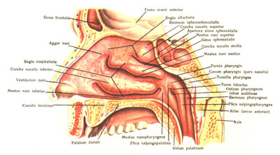
Fig. 88. Lateral wall of the nasal cavity, nasal conchae and passages. View from the right and inside (2/3).
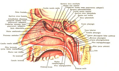
Fig. 89. The lateral wall of the nasal cavity and nasal passages with opening in them the anterior, middle and posterior cells of the ethmoid sinuses, the maxillary and frontal sinuses and nasolacrimal canal. View from the right and inside (2/3).
Lower, middle and upper turbinates removed.
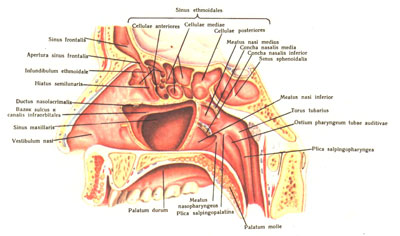
Fig. 90. Near-nasal sinuses. View from the right and inside (2/3).
The near-nasal sinuses are opened after removing a part of the lateral wall of the nasal cavity.
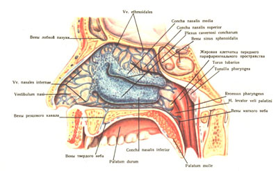
Fig. 91. Veins and plexus cavernosi concharum of the lateral wall of the nasal cavity. View from the right and inside (2/3).
Only the mucosa is removed.
The most complex structure is the lateral wall of the nasal cavity, which consists of: the nasal bone, the nasal surface and the frontal process of the upper jaw, the lacrimal and ethmoid bones, the perpendicular plate of the palatal bone and the medial plate of the pterygoid process. Three shells standing in the nasal cavity are located on the lateral wall. The upper and middle shells (concha nasalis superior and media) are part of the ethmoid bone, the lower (concha nasalis inferior) is an independent bone. The three shells correspond to the three nasal passages: the lower, middle and upper. The lower nasal passage (meatus nasi inferior) is located between the lower shell and the lower wall of the nasal cavity. In front of it opens the tear duct. The middle nasal passage (meatus nasi medius) is located between the lower and middle shells. Within the limits of the stroke, after removal of the middle turbinate, the lunate furrow (hiatus semilunaris) opens, described by N. I. Pirogov and called by him the oblique hemi-channel. Starting in the anterior-upper part of the nasal cavity by the extension (infundibulum ethmoi-dale), the sulcus is directed down and back, being located above and posterior to the processus uncinatus and below and anterior to the bulla ethmoi-dalis. An aperture (apertura sinus frontalis) opens in the anteroposterior funnel-like part of the furrow, leading to frontal sinus. The posterior low furrow also has an extension, at the bottom of which there is a hole (hiatus maxillaris) leading into the maxillary sinus. In addition, in the middle nasal passage and the lunate furrow, the anterior and part of the middle sinus ethmoidales cells open.
Upper bow (meatus nasi superior) is half as long as the middle one and is located between the middle and upper shells. In it and in the recessus spheno-ethmoidalis, the main sinus and part of the middle and posterior cells of the sinus ethmoidales open with a hole (apertura sinus sphenoidalis). The foramen sphenopalatinum is located behind and at the level of the upper nasal passage, hidden under the mucous membrane, connecting the nasal cavity with the pterygopalatomy.
Medially, all three nasal passages open into a common bowwhich is enclosed between the nasal septum and the nasal conchs.
The mucous membrane of the nasal cavity, covering the skeleton of the bone, repeats its relief and is divided into two areas in its structure and functions: the larger - respiratory (regio respiratoria) and the smaller - olfactory (regio olfactoria). The respiratory region captures the two lower nasal passages and the lower part of the middle shell. The olfactory region of the nasal mucosa contains special olfactory cells, the central processes of which are in the form of nn. olfactorii through lamina cribrosa penetrate into the anterior cranial hole and enter the bulb us olfactorius. The olfactory region occupies a small portion of the upper part of the nasal cavity, is distinguished by a yellowish color and extends to the upper nasal passage and the corresponding portion of the nasal septum. Under the mucosa are vessels and nerves.
The veins of the walls of the nasal cavity form a plexus that lies more superficially than the arteries and is especially well pronounced in the inferior and middle turbinates, where they resemble cavernous structures (plexus cavernosi concharum). The venous outflow from the plexus goes through the arteries accompanying the arteries, so that
the posterior walls of the nasal cavity bleed into the pterygoid plexus, from the upper sections into the veins of the orbit and the cavernous sinus, from the anterior sections along the veins around the anterior cartilage of the nose into the veins of the back of the nose, and then into the facial vein. In addition, the veins of the walls of the nasal cavity anastomose with the veins of the soft palate, pharynx and veins of the dura mater.
The main artery of the walls of the nasal cavity is the branch a. maxillaris - the main nebulae artery (a. sphenopalatina), which begins in the pterygopalatine fossa and through the same hole penetrates into the submucosal layer of the walls of the nasal cavity, where it is divided into aa. nasales posteriores laterales and a. nasalis posterior septi.
The upper wall, the upper sections of the lateral and medial walls of the nasal cavity, and the cells of the ethmoid bone are supplied by the anterior and posterior ethmoid arteries (branches of the orbital artery).
All the arteries supplying the walls of the nasal cavity, repeatedly anastomose with each other, with large palatine arteries, and in the region of the nasal vestibule with the branches of the facial artery.
Lymphatic vessels from the vestibule of the nasal cavity are anterior to the pear-shaped hole, penetrating between the edge of the hole and cartilage, or bend around the edge of the nostrils. Having penetrated into the subcutaneous tissue of the face, the vessels are sent to the submandibular or superficial parotid the lymph nodes. The lymphatic vessels of the posterior nasal cavity, perforating the side wall of the pharynx, are sent to the pharyngeal and upper deep cervical lymph nodes, anastomosing and merging with the lymphatic vessels of the palate, palatine tonsil, tongue and nasal and oral pharynx during its course.
Nerves. In the olfactory region nn spread. olfactorii. The mucous membrane of the remaining sections of the nasal cavity is innervated by the first and second branches trigeminal nerve. Nerves of the first branch: n. ethmoidalis posterior penetrates the hole of the same name and innervates the posterior cells of the ethmoid bone and the main collar; n. ethmoidalis anterior is the same as the artery of the same name, and divided into he. nasales laterales and mediales, innervates the upper and anterior part of the septum and the lateral wall of the nasal cavity, as well as the frontal sinus and ethmoid cell. Nerves of the second branch - he. nasales posteriores superiores laterales and mediales come from both the branch itself and ganglion pterygopalatinum.
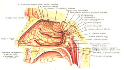
Fig. 92. Arteries and nerves of the lateral wall of the nasal cavity. View from the right and inside (2/3)
Veins and cavernous plexuses of shells were removed, the pterygo-channel was opened; prepared arteries and nerves.
They penetrate through the foramen sphe-nopalatinum and spread - the first in the area of the upper and middle shells, the second in the area of the back and bottom of the nasal septum. The largest of the medial branches is n. nasopalatinus - reaches the bottom wall, where it penetrates through the incisal canal to the anterior section of the hard palate mucosa, which innervates. Rr. nasales posteriores inferiores laterales depart from the anterior palatine nerve in the major palatal canal, pierce the bone and spread to the lower and middle nasal passages, the inferior nasal conch and the lower wall of the nasal cavity.
Related materials:

Popular
- Breast cancer is curable at any stage.
- The remedy for the cold Sinupret
- Azitrox - official instructions for use
- Chicken-bjaka: allowed antibiotics were found in Russian chicken
- Oral Cancer: Symptoms and Treatment
- Dark and thick blood during menstruation.
- Modern analogues of doxycycline tablets
- Is it possible to die from pneumonia
- What earwax will tell all about your health
- Tussin: instructions for use