Nose - Wikipedia. The structure of the human nose: anatomical features.
A person's nose is an organ of respiration and smell. In women, it is usually wider than in men, but, as a rule, shorter. Common building nasal cavity has no gender differences. A person’s nose performs the following functions: heats air currents from outside, retards the penetration of dust and microbes into the lungs, resonates the voice, and is directly involved in the discernment of odors.
In order to properly imagine the disease of the nose, you need to know its structure. The nose is the initial section of the upper respiratory tract.
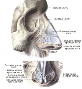
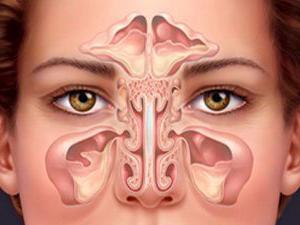
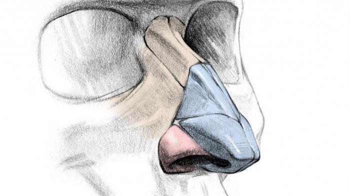
The anatomy of the human nose is as follows:external nose and the nasal cavity along with the paranasal sinuses. The external nose, having the appearance of an irregular trihedral pyramid, consists of cartilaginous, bony and soft parts. Its upper end, starting from the forehead, is the root of the nose; down and in front of it is the back of the nose, ending with the so-called tailbone of the nose. The structure of the wings of the nose is represented by the lateral convex and movable surfaces of the nose. Their lower free edges form the nostrils.
Human Anatomy: Nose Bones
The story about the structure of the nose and sinuses should begin with its location. From above, the nasal cavity borders on the cranial cavity, from top to bottom - oral cavity, and on the sides - with eye sockets. The nasal septum divides the cavity in half. Each half opens out nostrils. The nasal cavity is communicated posteriorly with the upper part of the pharynx by means of two oval shaped posterior nasal openings located next to each other, called Hoan.
Look at the photo of the structure of the nose:the posterior-upper bony part of the nasal septum consists of the vomer and the perpendicular plate of the ethmoid bone, and the anterior cartilaginous, of the quadrangular cartilage.
The outer wall of the nasal cavity, also called the side, is the most complex. Its structure includes nasal bone, as well as the frontal process and the nasal surface of the upper jaw body, the palatine bone, the ethmoid bone, the lacrimal bone, the pterygoid processes of the main bone.
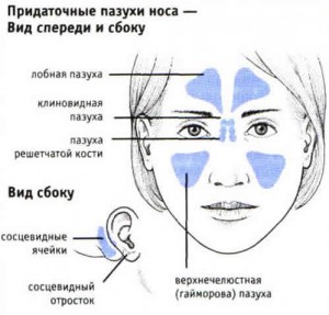
On the outer wall of the nasal cavity are three shells that divide the nasal cavity into the upper, middle and lower nasal passages. Under the lower shell is the opening of the nasal duct. Through special openings in the middle nasal passage, the paranasal sinuses open. The largest of them is maxillary, or maxillary. It is located in the body of the upper jaw.
Frontal sinus and anterior cells of the ethmoid labyrinth are located in the scales frontal bone. The posterior cells of the ethmoid labyrinth, as well as the main sinus, have a connection with the upper nasal passage.
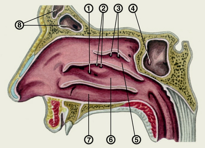
The sieve plate of the ethmoid bone in the anatomy of the nose forms the so-called roof of the nasal cavity. Its anterior slope is formed by the nasal bones, and the posterior by the anteroposterior wall of the main sinus.
The floor of the nasal cavity in the anterior part is composed of the palatal processes of the upper jaw, and in the posterior part it consists of horizontal plates of the palatal bones. The entire nasal cavity is lined with mucous membrane, which is covered with a multi-layered cylindrical ciliated epithelium. The movement of the hairs is directed backwards to the hoans.
Nose mucosa
Speaking about the structure of the nose of the person, special attention should be paid to the mucous membrane of the upper nasal passage. Together with the adjacent areas of the nasal septum mucosa and the upper part of the middle shell, it is lined with a specific sensitive epithelium. In it the peripheral endings of the branches of the olfactory nerve branch. This area of the mucous membrane is called the olfactory region. The rest of the nasal mucosa is called the respiratory region. It is lined with multi-layered cylindrical epithelium.
The thickness of the nasal mucosa in different areas is different. The most thin and poor mucous glands is the mucous membrane sinuses. The thickest is the shell mucosa. Due to the abundance of dense venous networks in the submucosal layer, cavernous or cavernous tissue is formed in places. It is most developed in the lower turbinate, as well as along the edge of the middle and at the posterior ends of the lower and middle cavities.
Various curvatures of the nasal septum, as well as other pathologies that develop in the nasal cavity, lead to the emergence of various diseases.
Article read 47 275 times (a).
The nose is an organ that is viewed more often from an aesthetic point of view, but its functions and significance for a person are much broader. It is especially important to disassemble the structure of the nose to understand the processes, both physiological and pathological, occurring in it.
Organ anatomy
Nasal cavity
Its appearance is different for different people, but its nose is the same in structure for everyone. There may be only minor deviations due to developmental features or injuries. Anatomically, the olfactory organ can be divided into three parts:
- the external nose is what we see on the face of a person;
- the inner part is a cavity, hidden from the eyes, in which the main work takes place;
- paranasal sinuses - voids in the paranasal space involved in the process of air exchange.
Now consider the structure of the nose of a person in more detail.
The outer part
It is formed by the bones of the skull, cartilage plates, muscle tissue and skin. A mathematician would compare an external nose with a three-sided irregular pyramid:
- the tip is called the nose and is located between the eyebrows;
- the back is the surface of the nose, formed at the junction of the two lateral bones, fastened by their upper edge to the root;
- the cartilaginous part continues the bone skeleton, forming the tip of the nose and its wings;
- the structure of the wings, in addition to cartilage tissue, includes the muscular structure, people do not use these muscles actively, they are mainly defined as mimic muscles, reflecting the emotional mood;
- the skin that is present in the structure of the nose is thin, but it is abundantly supplied with a network of capillaries, glands, richly innervated;
- the tip of the nose smoothly passes into the septum that separates and forms the nostrils - columella.
In front of the nose on the cartilaginous septum is the so-called Kisselbach zone - the place of the cluster blood vessels. Its close location leads to frequent nosebleeds. The outer part, especially the tip of the nose, is abundantly equipped nerve endings. Therefore, even the slightest injury in this area cause severe pain.
The formation of the internal structure of the nose ends no later than 13 years. It is believed that the final formation of the appearance ends by 15-16 years. But according to some sources, the human nose “matures” with the owner and changes throughout the person’s life.
Internal cavity
Nasal cavity
Anatomy and physiology of the nose implies, first of all, its internal structure, since it is here that all vital processes occur.
The cavity of the olfactory organ has its own borders, which are formed by the bones of the skull, the eye sockets, the oral cavity, separated by the sky. "Entry gates" for circulating air are nostrils. In the back part of the structure of the internal cavity forms two holes in the upper half of the pharynx, called choans.
The septum consists of cranial bones and a cartilaginous plate; it divides the internal cavity into two approximately equal, half - nasal passages.
In the structure of each of them there are five walls:
- the lower part is represented by the maxillary bones constituting the sky;
- the top wall is otherwise called perforated, since its structure has openings for the passage of large blood supplying vessels and branching of the nerve trunk;
- the inner (medial) wall is a septum of the nose, consists of bone and cartilage tissue;
- the structure of the outer (lateral) side is rather complicated, here are the nasal conchae - the formation of the ethmoid bone: the upper, middle, lower and corresponding respiratory passages. The upper two passages have access to the frontal sinuses, which explains the possibility of the development of frontal sinusitis in complicated infections in the nose. But the lower respiratory passage is an anatomical conductor of the lacrimal secretion into the cavity, since ducts of the lacrimal glands exit into it.
The structure of the internal cavity implies the division into three zones, each of which is distinguished by its structure:
- The vestibule is a zone within the wings of the nose that is lined with epithelial cells and contains many fine hairs.
- The respiratory zone includes the lower nasal passage; goblet cells produce mucus secretion in the cell structure. Layer below is submucous tissue, containing abundant blood, lymphatic vessels, ducts of glands, nerve endings.
- The sense of smell is the space that encompasses the middle and upper shell of the nose. Internal structure represented by epithelial cells with many olfactory cilia.
Sinus location
It is impossible to explain how the nose of a person works without telling about the paranasal sinuses. Sinuses are essentially free cavities involved in all processes in which the nose itself is involved.
For the treatment and disposal of MOKROTY our readers successfully use a natural remedy for MOKROTHA. This is 100% natural remedyThe basis of which includes only herbs, and mixed in such a way as to most effectively deal with the disease. The product will help to quickly and effectively defeat the cough in a short time, and once and for all. Since the drug consists only of herbs, it does not have any side effects. Does not affect pressure and heart rate. Get rid of sputum ... "
Deeper to understand the construction of the nose will help the scheme, with the full designation of the internal cavity and sinuses.
The functional value of the body
It would seem a strange question, why does a person need a nose, but everyone will be able to answer exactly what the significance of this body is. In the meantime, it performs several important functions, which accounts for its complex structure.
Ensuring comfortable breathing
The main task of the nose - ensuring normal air circulation in sufficient quantities. It is known that cold air is poorly absorbed by the body. The anatomy of a person’s nose is such that when it enters the cavity, cold streams, passing through passages richly equipped with blood vessels, warm up to the temperature of comfort. Due to the unique structure, the nose is involved in the process of breathing as a primary and important link.
The unfavorable picture is visible in violation of normal breathing through the nose, which can be caused by an inflammatory process, allergic edema or changes in the structure of the partition. At the same time, breathing takes place predominantly through the mouth, cold streams enter the throat and larynx which are not adapted for this.
Most dangerous chronic rhinitis. In this case, the functions of the nose are partially lost, the body suffers from lack of oxygen, which leads to various pathological processes in many organs and systems: vascular, cardiac, nervous.
Odor difference
The nose, as a sense organ, refers to the sense of smell, that is, to the ability to discern the smallest odors. This is due to the special structure and the presence of ciliated epithelial cells that line the top two shells. The cilia of these cells are able to catch and recognize even a small amount of the substance that enters the nose.
The ciliated epithelium is associated with the olfactory nerve, the signal coming from the outside through the nerve is addressed to the brain, analyzed and defined as pleasant or repulsive, dangerous or favorable.
Smell and touch complement each other. We observe this process when, with a strong nasal congestion, taste differences disappear, and the food becomes equally fresh.
Protective function
Nose structure
What is the nose for, except for the above functions? Protecting the body from the penetration of foreign particles and pathogenic bacteria is one of the main tasks of the nose, due to its structure.
The first barrier on the way foreign bodies becomes the threshold. The tip of the nose is provided with hairs that retain large particles. Deeper microparticles and harmful microorganisms are affected by nasal mucus, which has a bactericidal effect. The greater the number of pathogenic microflora, the more irritation occurs. trigeminal nerveincreases secretion. The act of sneezing is a way to release the nose from accumulated particles.
Reflex response, due to the structure of the nose, can be called and lacrimation with the penetration of the virus into its cavity or sharp irritating odors.
Participation in sound
Surprisingly, in the formation of speech plays a significant role nose and its anatomy allows you to amplify the sounds of different frequencies. This is particularly evident in the pronunciation of consonants. The change in sound formation is observed when severe edema nasal mucosa and sinuses, which is heard as nasal. Moreover, the structure of this body determines the unique timbre of the individual's voice.
Why do we need paranasal sinuses
Paranasal sinuses are a very important and necessary element of the nose, the structure of which defines the following functions:
- warming the incoming air flow and moistening it;
- participation in the organization of the sense of smell;
- the presence of free cavities reduces the weight of the skull, respectively, reduces the load on the spinal column;
- mitigate impact force in injuries;
- given the structure, are involved in the regulation of voice timbre.
Features of development in children
In newborns, the structure of the olfactory organ is very different from adults. The shells are underdeveloped, the external nose is small, consisting of the same sections as the adult. The strokes of the olfactory organ are narrow, which contributes to the development of frequent inflammatory processes. At the same time, the structure of the nasal mucosa is marked by a saturated circulatory grid, which causes rapid edema during hypothermia.
The child's sense of smell is not able to perform its functions in full, the possibility of warming the air during breathing is formed only by five years. Therefore, in young children at temperatures below -5–10 degrees, the tip of the nose quickly freezes.
The paranasal sinuses have a different structure. A baby is born, having in its infancy maxillary sinuses and lattice labyrinth cells. With age, the structure of the labyrinth changes, its volume increases. The final formation of the maxillary cavities ends closer to 12 years. And the frontal sinuses and the sphenoid at the age of 3-5 years only begin their construction.
The air, inhaled by man from the environment, before getting into the lungs, must be heated and cleaned from dust and other microparticles. This function is performed by the human nose, which has its own structural features, which determines the functionality of the nasopharynx. The correct structure of the nose plays a big role in the relationship of the human body with the environment.
What does the nose consist of?
The anatomy of the nose is quite simple, this organ consists of the external part and the nasal cavity. It performs many functions - defensive, resonant, olfactory, and others.
Outdoor department
The external part of the nose consists of two bones, it is from them that the upper back of this organ is formed, its lower part consists of the cartilages that form the basis of the wings and the tip of the nose. Sometimes, however, this organ can have a slightly different structure, it can only be determined with the help of MRI of the nose.
Based on the fact that the nasal sinuses are located deep enough, to explore them by some other method fails. In the process of MRI, changes in the structure and tissues in the early stages of the development of pathologies are detected.
Cartilage form a pair of side walls, wings of the nose, nostrils, nasal septum. The bones and cartilages that make up the outer part of the organ are covered on top of the skin, consisting of sebaceous glands, capillaries and nerve fibers. From the two sides of the wings of the nose are holes - nostrils, it is through them that the air enters the lungs.
Around the nose are the paranasal sinuses, they are necessarily closely connected with the nasal cavity. There is a classification according to which four pneumatic sinuses are distinguished - maxillary, cells of the ethmoid labyrinth, frontal, wedge-shaped. An MRI of the sinuses allows specialists to conduct a deep examination of all the sinuses with maximum precision. With the help of such a diagnosis, pathologies can be identified at an early stage of their development or else they can be prevented.
 In addition, the paranasal sinuses are divided into front and rear. The division of these components of the human nose is convenient, first of all, for doctors in that the pathologies of the anterior and posterior sinuses are different. Using MRI of the sinuses, these differences can be traced, however, in the case of the occurrence of pathological phenomena in the paranasal sinuses. As medical practice shows, diseases of the posterior sinuses are much less common than those of the front.
It is important to know some features of the structure of the nose of a person immediately after birth and at the stage of formation of this organ of the respiratory system. It is known that children have only two sinuses - the maxillary and ethmoid labyrinth. However, these paranasal sinuses are represented by rudiments; they are at the stage of development. In children, all the nasal passages are significantly narrower than in adults, which accounts for the periodic observation of difficulty breathing in infants.
Nasal cavity performs the main function - air purification from dust and foreign particles. At the entrance to it are small hairs that perform a protective function. The structure of the nose provides reliable protection of the respiratory tract, as it protects the body also mucus, which is secreted by the goblet glands.
In the absence of pathologies, the mucus of the goblet glands is endowed with antiseptic properties, due to which it is capable of destroying the pathogenic bacteria that enter the nasal cavity. In addition, this mucus eliminates the likelihood of too cold and dry air in the body.The nasal cavity consists of four walls:
- lower;
- top;
- medial;
- lateral.
 The anatomy of the nose includes another small area consisting of many blood vessels, for this reason, nosebleeds are often found in this area. The bone and cartilaginous septum divides the nasal cavity into two approximately equal parts, in some cases, when receiving an injury or in the process of voluminous formations, a curvature of the nasal septum may occur, which usually violates breathing.
In case of any violations, magnetic resonance imaging will help to detect pathological processes, with its help a specialist can notice any changes in the structure of the nasopharynx. In cases where the doctor cannot see all the sinuses, additional diagnostic methods such as x-rays or computed tomography can be used.
If you have questions to the doctor, please ask them on the consultation page. To do this, click on the button:
Related records

Clinical anatomy of the external nose
Hoc (nasus) consists of an external nose and nasal cavity.
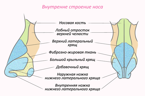
The external nose (nasus externus) is represented by a bone-cartilaginous skeleton in the shape of a pyramid (Fig. 1.1), covered with skin. It distinguishes the tip, root (nose), back, rays and wings.
Bone part of the skeletonconsists of paired flat nasal bones and frontal processes of the upper jaw. These bones, together with the anterior nasal spine, form a pear-shaped opening in the facial skeleton. Cartilaginous part of the skeletonconsists of paired triangular and winged, as well as additional cartilage; the wings of the nose in its lower back part lack cartilage base. The skin in the lower third of the nose has many sebaceous glands. Bending over the edge of the entrance to the nose (nostrils), it lines the walls of the vestibule of the nose (vestibulum nasi) for 4-5 mm. Here on the skin there is a large amount of hair, which makes it possible for boils and sycosis to occur. In the area of the wings of the nose under the skin are the muscles that expand and constrict the entrance to the nose.
The external nose, as well as all the soft tissues of the face, is characterized by abundant blood supply: there are branches that anastomose among themselves from the maxillary and orbital arteries, from the external and internal carotid arteries, respectively. The veins of the external nose drain blood through the anterior facial vein into the internal jugular vein and in large quantities through the veins of the nasal cavity, then through the orbital veins into the venous plexus of the pterygopulmonary fossa (plexus pterygoideus) and into the cavernous sinus (sinus caver-nosus), middle medulla ( v.meningea media) and then into the internal jugular (v.jugularis interna) veins.
Lymphatic drainage from the external nose is carried out mainly in the submandibular lymph nodes. Muscles of the external nose are innervated by sprigs. facial nerve (n.facialis), the skin is the first (optic nerve - n.ophtalmicus) and the second (maxillary nerve - n.maxillaris) branches of the trigeminal nerve, supraorbital (n.supraorbitalis) and infraorbital (n.infraorbitalis) nerves.
The plastic skin-cartilaginous structure of the anterior part of the external nose allows within certain limits to shift it to the sides without subsequent persistent deformation. However, a strong mechanical effect on the osseous part of the nose is often accompanied by fractures of the nasal bones, often with displacement of fragments, and with a more severe injury, a fracture of the frontal processes of the upper jaw.
Clinical anatomy of the nasal cavity
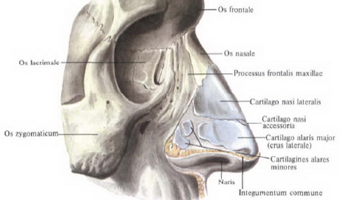
The nasal cavity (cavum nasi) is located between the cavitymouthand anterior cranial fossa,and from the sides - betweenpaired upper jawsand paired lattice bonemi.The nasal septum divides the sagittal into two halves, opening anteriorly with the nostrils and posteriorly, into the nasopharynx, with the joans. Each half of the nose is surrounded by four airway paranasal sinuses: maxillary,lattice labyrinth, frontal and wedge-shaped,which communicate on their side with the nasal cavity (Fig. 1.2). The nasal cavity has four walls: lower, upper, medial and lateral; posterior nasal cavity through Joan communicates with the nasopharynx, the front remains open and communicates with the outside air through the holes (nostrils).
Lower wall (bottom of the nasal cavity)formed by two palatal processes of the upper jaw and in a small area of the posterior part - by two horizontal plates of the palatine bone (hard palate). By a similar line, these bones are connected by a suture. Violations of this compound lead to various defects (cleft of the palate, cleft lip). In the front and in the middle in the bottom of the nasal cavity there is a nasal channel (canalis incisivus), through which the same-name nerve and artery anastomosing in the canal with the large palatine artery pass into the oral cavity. This circumstance must be kept in mind when performing submucous resection of the nasal septum and other operations in this area in order to avoid significant bleeding. In newborns, the bottom of the nasal cavity is in contact with the tooth buds, which are located in the body of the upper jaw.
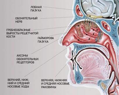
Upper wall (roof)the nasal cavity in the front is formed by the nasal bones, in the middle sections by the ethmoid plate (lamina cribrosa) and the ethmoid cells (the largest part of the roof), the back sections are formed by the front wall of the sphenoid sinus. Threads of the olfactory nerve pass through the holes in the ethmoid plate; the bulb of this nerve lies on the cranial surface of the ethmoid plate. It must be borne in mind that in the newborn lamina cribrosa is a fibrous formation that ossifies only by 3 years.
Medial wall,or nasal septum(septum nasi), consists of the anterior cartilaginous and posterior bone sections (Fig. 1.3). The bone section is formed by the perpendicular plate (lamina perpendicularis) of the ethmoid bone and vomer (vomer), the cartilaginous - quadrangular cartilage, the upper edge of which forms the anterior part of the back of the nose. On the eve of the nose, anteriorly and downwards from the front edge of the quadrangular cartilage, there is an outside skin-webbed mobile part of the nasal septum (septum mobile). In a newborn, the perpendicular plate of the ethmoid bone is represented by a membranous formation, the ossification of which ends only by 6 years. The nasal septum is usually not exactly in the median plane. Significant curvatures of her in the anterior section, more common in men, can cause disturbances in breathing through the nose. It should be noted that in a newborn the height of the opener is less than the width of the choana, therefore it appears as a transverse slit; only by the age of 14, the height of the opener becomes greater than the width of the choana, and it takes the form of an oval that extends upwards.
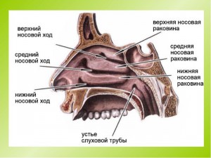
Structure lateral (external) wall of the nasal cavitymore complicated (Fig. 1.4). In its formation take part in the front and middle parts medial walland frontalappendix of the upper jaw, lacrimaland medial nasal bonesurfacethe ethmoid bone, in the posterior part, forming the edges of the choana, is the perpendicular process of the palatine bone and the wing-palatal processes of the sphenoid bone. On the outer (lateral) wall are located three bow conchs(conchae nasales): lower (concha inferior), medium (concha media) and upper (concha superior). The lower shell is an independent bone, the line of its attachment forms an arc, convex upwards, which should be considered when puncturing the maxillary sinus and conchotomy. The middle and upper shells are processes of the ethmoid bone. Often the front end of the middle shell is inflated in the form of a bubble (conhae bullosa) - this is an air-cell of the ethmoid labyrinth. Anterior to the middle shell there is a vertical bone protrusion (agger nasi), which can be expressed to a greater or lesser extent. All the nasal conchs, attaching with one lateral edge to the lateral wall of the nose in the form of oblong flattened formations, with the other edge hang downwards and medially in such a way that under them are formed according tovenous lower, middle and upper nasal passages,which height is 2-3 mm. The small space between the upper sink and the roof of the nose, called sphenoemoidal, is usually referred to as the upper nasal passage. Between the nasal septum and the nasal concha remains a free space in the form of a slit (3-4 mm in size), which extends from the bottom to the roof of the nose - a common nasal passage.
In a newborn, the lower conch descends to the bottom of the nose, the relative narrowness of all nasal passages is noted, which causes a rapid onset of difficulty in nasal breathing in young children, even with a slight swelling of the mucous membrane due to its catarrhal state.
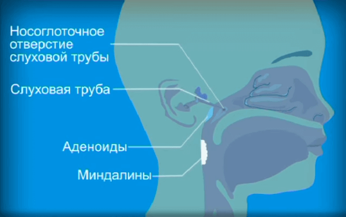
On side wall of the lower nasal passageat a distance of 1 cm in children and 1.5 cm in adults from the front end of the sink is the excretory nasolacrimal opening.This hole is formed after birth; in case of a delay in its opening, the outflow of tear fluid is disturbed, which leads to cystic dilation of the canal and narrowing of the nasal passages. The bone of the lateral wall of the lower nasal passage at the base is much thicker than that of the line of attachment of the lower shell (this must be borne in mind when puncturing the maxillary sinus). The posterior ends of the lower cavities closely fit the pharyngeal orifices of the auditory (Eustachian) tubes on the lateral walls of the pharynx, as a result of which the hypertrophy of the shells may impair function. auditory tubes and develop their disease.
Middle nasal passagelocated between the lower and middle shells, on its lateral wall is the crescent (semi-lunar) gap (hiatus semilunaris), the posterior section of which is located below the front (first described by N.I. Pirogov). This slot opens: in the posterior part - the maxillary sinus through the hole (ostium 1 maxil-lare), in the anteroparticular - the opening of the frontal sinus canal, which does not form a straight line, which must be kept in mind when probing the frontal sinus. The crescent-shaped gap in the posterior part is limited by the protrusion of the ethmoidal labyrinth (bulla ethmoidalis), and in the anterior region by the hooked process (processus uncinatus), which departs anterior to the anterior edge of the middle turbinate. The medial nasal passage also opens the anterior and middle cells of the ethmoid bone.
Upper nasal passageextends from the middle shell to the roof of the nose and includes sphenoemoidal space. At the level of the posterior end of the upper shell, the sphenoid sinus is opened through the opening (ostium sphenoidale) into the upper nasal passage. The posterior cells of the ethmoid labyrinth also communicate with the superior nasal passage.
Nasal mucosacovers all its walls with a continuous layer and continues into the paranasal sinuses, throat and middle ear; she is does not have a submucosal layer thatry generally absent in the respiratory tract, with the exception of the subglossal region of the larynx.The nasal cavity can be divided into two sections: the front - nasal vestibule(vestibulum nasi) and proper nasal cavity(cavum nasi). The latter, in turn, is divided into two areas: respiratoryand olfactory.
The respiratory region of the nasal cavity (regio respiratoria) occupies the space from the bottom of the nose up to the level of the lower edge of the middle shell. In this area mucousthe shell is covered with a multi-row cylindrical ciliatedepithelium.
Under the epithelium is the actual tissue of the mucous membrane (tunica propria), consisting of connective tissue collagen and elastic fibers. There is a large number goblet cells that produce mucus, andtubular-alveolar branched glands producingserous or serous-mucous secretion that through excretoryducts coming to the surface of the mucous membrane.Slightly below these cells, basal cells are located on the basement membrane, which are not subject to desquamation. They are the basis for the regeneration of the epithelium after its physiological and pathological desquamation (Fig. 1.5).
The mucous membrane is tightly welded all the way to the perichellate or periosteum that makes up single wholetherefore, during the operation, the membrane is separated with these formations. In the region of the predominantly medial and lower parts of the lower shell, the free edge of the middle shell and their hind ends, the mucous membrane is thickened due to the presence cavernous tissueconsisting of dilated venous vessels, the walls of which are richly supplied with smooth muscles and connective tissue fibers. Portions of the cavernous tissue can sometimes be found on the nasal septum, especially in its posterior part. Filling and emptying the cavernous tissue with blood occurs reflexively under the influence of various physical, chemical and psychogenic stimuli. The mucous membrane containing cavernous tissue can instantly swell (thereby increasing the surface and warming the air to a greater extent), causing a narrowing of the nasal passages, or shrinking, having a regulating effect on the respiratory function. In children, the cavernous venous formations reach their full development by the age of six. At a younger age, rudiments of the Jacobson olfactory organ are sometimes found in the mucous membrane of the nasal septum, located at a distance of 2 cm from the front edge of the septum and 1.5 cm from the bottom of the nose. Here cysts can form and develop inflammatory processes.
The olfactory region of the nasal cavity (gegio olfactoria) is located in its upper sections, from the arch to the lower edge of the middle turbinate. In this area, the mucous membrane covers olfactory epithelium,the total area of which in one half of the nose is about 24 cm 2. Among the olfactory epithelium in the form of islets, there is a ciliated epithelium, which performs a cleaning function here. The olfactory epithelium is represented by olfactory fusiform, basal and supporting cells. The central fibers of the spindle-shaped (specific) cells pass directly into the nerve fiber (fila olfactoria); the tops of these cells have protrusions into the nasal cavity - olfactory hairs. Thus, the spindle-shaped olfactory nerve cell is both a receptor and a conductor. Surfacethe olfactory epithelium is covered by the secret of specific tubeschato-alveolar olfactory (bowman) glands, whichis a universal solvent of organic substances.
The blood supply to the nasal cavity (Fig. 1.6, a) is provided by the terminal branch of the internal carotid artery (a.ophthalmica), which in the orbit extends the lattice arteries (aa.ethmoidales anterior et posterior); these arteries feed the anteroposterior sections of the walls of the nasal cavity and the ethmoid labyrinth. The largest artery of the nasal cavity- a. sphenopalatina (branch of the internal maxillary artery from the systemexternal carotid artery),it comes out of the pterygopalatine fossa through the hole formed by the processes of the vertical plate of the palatine bone and the body of the main bone (foramen sphenopalatinum) (Fig. 1.6, b), gives the nasal branches to the side wall of the nasal cavity, septum and all the paranasal sinuses. This artery is projected on the side of the nose near the posterior ends of the middle and lower turbinates, which must be borne in mind when performing operations in this area. Feature of vascularization of the nasal septumis the formation of a dense vascular network in the mucous membrane in the region of its anterior third (locus Kisselbachii), here the mucous membrane is often thinned (Fig. 1.6, c). Nasal bleeding occurs more often from this place than from other areas, so it is called the "bleeding area of the nose." Venous vessels accompany the arteries. A feature of the venous outflow from the nasal cavity is its connection with the venous plexuses (plexus pterigoideus, sinus cavernosus), through which the nasal veins communicate with the veins of the skull, orbit and pharynx, as a result of which it is possible to spread the infection along these routes and the occurrence of rhinogenic intracranial and orbital complications, sepsis and others
Lymphatic drainage from the anterior sections of the nose is carried out in the submandibular the lymph nodes, from the middle and posterior parts - into the deep cervical. It is important to note the connection of the lymphatic system of the olfactory region of the nose with the intershell spaces, carried out along the perineural paths of the olfactory nerve fibers. This explains the possibility of meningitis after an operation on the lattice labyrinth.
In the nasal cavity, olfactory, sensitive and secretory innervation is distinguished. Olfactory fibers (fila olfactoria) diverge from the olfactory epithelium and through the ethmoid plate penetrate the cranial cavity to the olfactory bulb, where they form synapses with the dendrite of the olfactory tract cells (olfactory nerve). The parahippocampal gyrus (gyrus hippocampi), or seahorse gyrus, is the primary center of smell, the hippocampal cortex (ammonium horns) and the perforative anterior substance are the highest anterior cortical center of smell.
Sensitive innervation of the nasal cavity is carried out by the first (n.ophtalmicus) and second (n.maxillaris) branches of the trigeminal nerve (Fig. 1.7). From the first branch of the trigeminal nerve, the anterior and posterior ethmoid nerves, which penetrate into the nasal cavity along with the vessels and innervate the lateral divisions and the nasal cavity, depart. The second branch is involved in the innervation of the nose directly and through the anastomosis with a prilatine node, from which the posterior nasal nerves extend mainly to the nasal septum. The infraorbital nerve departs from the second branch to the mucous membrane of the bottom of the nasal cavity and maxillary sinus. The branches of the trigeminal nerve anastomose among themselves, which explains the irradiation of pain from the nose and paranasal sinuses to the teeth, eyes, dura (pain in the forehead, occiput), etc. The sympathetic and parasympathetic innervation of the nose and paranasal sinuses is represented by the nerve of the pterygopathic canal (vidiy nerve), which originates from the plexus on the internal carotid artery (upper cervical sympathetic node) and the cranial assembly of the facial nerve (parasympathetic portion).
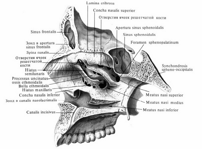
Clinical anatomy of the paranasal sinuses
The near-nasal sinuses are located around the nasal cavity and communicate with it (Fig. 1.8). Only four pairs of air sinuses: maxillary, ethmoid labyrinth cellsrint, frontaland wedge-shaped.There are anterior (maxillary, frontal, anterior and middle cells of the ethmoid bone) and posterior (sphenoid and posterior cells of the ethmoid bone) of the sinuses. Such a unit is convenient, since the pathology of the anterior sinuses is somewhat different from that of the posterior sinuses. In particular, front sinuses communicate with the cavitythe nose through the middle nasal passage, and the rear- through the top,what is important in diagnostic terms; diseases of the posterior sinuses, especially sphenoid, are much less common than the front.
Maxillary sinuses(sinus maxillaris) paired, located in the body of the upper jaw (see Fig. 1.8). They are the largest: the volume of each of them is on average equal to 10.5-17.7 cm 3 (from 1.5 to 31.5 cm). The inner surface of the sinuses is covered with a mucous membrane with a thickness of about 0.1 mm. The multi-row cylindrical ciliated epithelium covering the mucous membrane functions (has clearance) in such a way that the mucus moves upward in a circle to the medial angle of the sinus, where the fistula is located with the middle nasal passage of the nasal cavity. In the sinus distinguish anterior and posterior, upper and lower, as well as the medial wall.
On the front, or front, wall outside there is a groove - canine, or canine, fossa (fossa canina). It should be borne in mind that when palpating this wall through the soft tissue of the cheek immediately above the fossa, the infraorbital nerve (n.infraorbitalis) comes out of the bone. Canine fossa can be of different depths (average 4-7 mm). At its considerable depth, the anterior and upper walls of the sinus are in the immediate vicinity of the medial wall. In such cases, when the puncture of the sinus through the lower (and even more so through the middle) nasal passage, the needle imperceptibly for the surgeon can penetrate the anterior or upper wall into the soft tissues of the cheek or orbit, which can lead to the development of purulent complications. In the region of the canine fossa, the anterior wall is the thinnest.
The medial (nasal) wall of the sinus is bony, only in its upper section can there be no bone, and then in this place the wall is represented only by duplication of the mucous membrane. The medial wall corresponds to the lower and middle nasal passages. In its anterior section, there is a nasal duct, and in the upper, corresponding to the middle nasal passage, under the orbital rim there is a sinus opening in the nasal cavity (ostium maxillare). Sometimes there is not a simple hole, but a channel with a length of several millimeters. The location of the exit from the sinus in its upper section, its relative narrowness (diameter 2-6 mm) and in some cases the presence of not a hole, but a channel (or several holes - a fountain) creates unfavorable conditions for outflow of discharge from the sinus, which contributes to the development of inflammatory process. In the upper part, the medial wall of the sinus borders with the cells of the ethmoid bone, which often allows the inflammatory process to spread in this direction.
Upper wall maxillary sinus at the same time is the lower wall of the orbit; this wall is the thinnest, passing through the canal of the infraorbital nerve and the vessels of the same name; sometimes there are degiscences (congenital clefts in the bone), closed only by the mucous membrane. In this regard, during the operation, the contents of the orbit can be damaged through such degradations. In some cases, the upper and medial walls of the sinus are at a short distance from each other; in such conditions, puncture of the sinus through the nasal passage is dangerous, since the needle can penetrate the orbit and cause purulent inflammation in her.
The lower wall, or bottom, of the sinus is the alveolar process of the upper jaw; In most cases, in adults, the bottom of the sinus is below the bottom of the nasal cavity. It is important to note that in adults, the 2nd premolar and the 1st molar are closest to the bottom of the sinus, in some cases the tops of the roots of the teeth will stand in the sinus and are covered only by the mucous membrane. This explains the often observed spread of the inflammatory process from the corresponding teeth to the sinus.
The back wall of the sinus is thick, formed by the maxillary tubercle, which encloses the pterygopulla fossa in front, where the maxillary nerve is located, the pterygoid node, the internal maxillary artery, the pterygo-venous plexus.
Ethmoid sinusesor the ethmoidal labyrinth (labyrinthus ethmoidalis), is represented by the ethmoid pneumatic cells, which are located between the frontal and sphenoid sinuses (see Fig. 1.8). Outside, the ethmoid cells border the paper plate of the orbit, and the medial wall of the ethmoid bone is the lateral wall of the nasal cavity. The number, volume and location of the lattice cells vary; on average, they are 8-10 on each side. Frequently observed options for the location of lattice cells - their distribution in the orbit in the anterior or posterior sections. In this case, they are bordered at different lengths and with the anterior cranial fossa. Often there is also an option when the lattice labyrinth cells are located laterally to the lattice plate on both its sides; in these cases, the boundary between the cranial cavity and the nasal cavity are both the cribriform plate and the cribrius arch. At the same time, in surgical terms, it is important to note that the cribriform plate more often lies lower than the crib of the cribriform bone on each side of it, therefore, when opening the cage labyrinth cells, one should strictly adhere to the lateral direction so as not to penetrate the cranial cavity through the cage bone.
Frontal sinus(sinus frontalis) is in the scales of the frontal bone (Fig. 1.9). The sinus has four walls: anterior (facial), posterior (cerebral), bordering the cranial fossa, lower (orbital), most of which is the upper wall of the orbit and which for a short distance borders on the ethmoid bone cells and nasal cavity, and the medial (interphalar) ), which in the lower part is usually located in the middle line, and upwards it can deviate to the sides. Front and back wall in the upper section of the sinus converge at an angle. On the lower wall of the sinus anterior to the septum is the opening of the fronto-nasal canal, whose length is about 1 - 1.5 cm; In some cases, the sinus opens into the nasal cavity not with a channel, but with a hole. Usually, the canal opens in the anterior part of the lunate gap in the middle nasal passage. The configuration and dimensions of this sinus are variable, its volume is on average 4.7 cm 3. Sometimes one or both of the sinuses are missing, which is important in the diagnostic plan. In some cases, the sinuses, extending laterally, can be large, have bays and partitions.
Sphenoid sinuses(sinus sphenoidalis) are located in the body of the sphenoid bone (see Fig. 1.9). In each sinus distinguish anterior, posterior, upper, lower, outer and inner walls. Sinus divides interpusal septum, or inner wall. In the anterior wall of each sinus there is a discharge opening (ostium sphenoidale), leading to the upper nasal passage. Such communication of the sinus with the nasal cavity causes the outflow of the discharge into the nasopharynx along its back wall. The interbasal septum extends anteriorly to the nasal septum. The lower wall of the sinus partially forms the arch of the nasopharynx, the upper wall is represented by the lower surface of the Turkish saddle; besides the pituitary and the optic nerve, a portion of the frontal lobe of the brain with olfactory gyrus adjoins this wall above. The posterior wall is thicker and passes into the basilar region. occipital bone. The lateral wall of the sphenoid sinus is most often thin (1-2 mm), with it borders the internal carotid artery and cavernistaya sinus(sinus cavernosus); here are the oculomotor nerve, the first branch of the trigeminal, block and outlet nerves (III, IV, V, VI pairs of cranial nerves).
The newborn has only two pairs of sinuses - maxillary and ethmoid, however, and these sinuses are represented only by rudiments. Thus, the maxillary sinuses are only diverticula of the nasal mucosa in the thickness of the upper jaw at the inner corners of the sockets in the form of a slit 10 mm long, 2-3 mm wide and high. By the age of 6, these sinuses acquire normal forms, but their size is often small; by the age of 8, the bottom of the sinuses drops to the level of the bottom of the nose, and only by the age of 12, it is below the bottom of the nasal cavity, as in an adult. It is of interest to the clinic that in infancy the interrelations of the teeth, orbit and the maxillary sinus have significant features. If an adult has a sinus between the orbit and teeth, in an infant, the lower wall of the orbit is located directly above the two rows of rudiments of dairy and permanent teeth, and the germ of the sinus is medially at some distance from the teeth. With an increase in the child's age, the teeth gradually take up their permanent place, and the maxillary sinus takes on the appropriate dimensions and configuration. In early childhood, the canine is closest to the sinus; at the age of 6, there are two premolars and a molar near the bottom of the sinus, which for one reason or another can cause the disease of the maxillary sinus (as in an adult). By the age of 12, the topography of these formations is approaching the norm of an adult.
Cells of the ethmoid bone at the time of birth formed, but their number and volume increase with age, especially in the period from 3 to 5 years.
Frontal and sphenoid sinuses in a newborn are absent; their formation begins by 3-4 years. The wedge-shaped sinuses appear to be, as it were, laced cells of the ethmoid labyrinth, located in the body of the sphenoid bone. Frontal sinuses appear at the upper inner angle of the orbit from the anterior cells of the ethmoid bone; the nasal mucosa grows into them, while the spongy bone between the outer and inner cortical plates of the frontal bone continues to dissolve. At the age of 6 years, the height and width of these sinuses are about 8 and 12 mm, respectively; in some cases only one frontal sinus can form, sometimes they are both absent.
There are more people in the world who are thinking about the fact that they do not like the shape of their nose, than those who are thinking whether they can make them breathe better. Of course, everyone knows about everyday care, treatment for diseases, etc. But how many of us think about what the nasal cavity is?
Anatomy of the airways
Lung tissue is a rather delicate structure. That is why the air, before making the way to them, must be cleaned from dust and part of the microbes, humidified and warmed. This state of his is achieved with the help of a complex breathing apparatus having a complex structure.
Before reaching the lungs, the air passes through the trachea, above is the larynx and nasopharynx, as well as the upper part - the cavity where it falls immediately after inhalation. It is here that its primary processing takes place.
Nose structure
Few people think about it, but breathing provides us with a very perfect and complex organ. Perhaps that is why any, even small problems instantly affect the state of health. Conventionally, this body can be divided into two large parts:
- external nose;
- nasal cavity;
- paranasal sinuses.
The part that each person sees, simply by viewing his face in the mirror, is formed by small bones and cartilage tissue. Finally, its form is formed by about the 15th year of life.
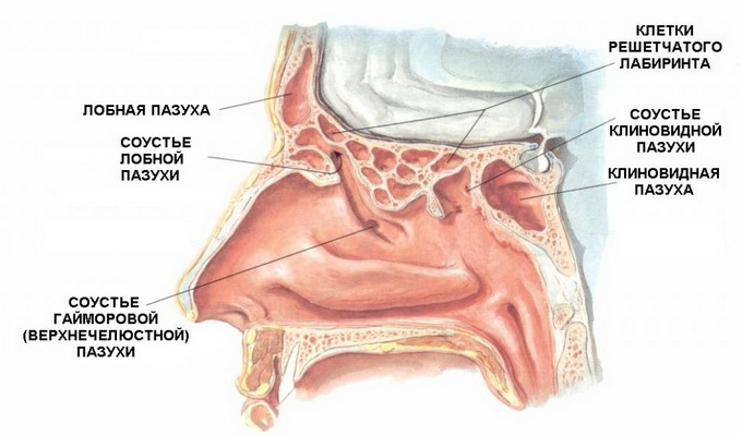
The structure of the nasal cavity is so difficult due to the fact that it is here that the regulation of the temperature of the inhaled air and its purification takes place. The vestibule is lined here; there are small hairs that trap particles of dust and germs. Three curved bone plates protrude into the cavity, which form the so-called shells. Some of their areas are lined with sensitive cells, thanks to which a person has a sense of smell. Here, through the narrow passages, the accessory sinuses - the maxillary, frontal, main and ethmoid - have access. What are they made of and why are they needed?

Nasal congestion
It would seem, why complicate things? Let the air simply pass into the lungs, let its path be brief and simple. But evolutionary development ordered otherwise, and man has not just a nose. The nasal cavity has four additional sinuses.
- Maxillary, or maxillary. This sinus is the most voluminous - up to 30 cubic centimeters. In shape, it resembles a tetrahedron. This cavity communicates with the main (main) through the course in the common wall. In the projection on the front of the face, these sinuses are located on the sides of the nose immediately under the eyes.
- Frontal This sinus, on the contrary, is very small - only 3-5 cubic centimeters. It is located in the frontal bone and also communicates with the nose through a narrow passage.
- Grid. These sinuses consist of separate bone cells, so they are sometimes called a labyrinth. These cavities are in a rather inaccessible place and border on the inside of the orbit and the brain.
- The main (main). This part is the least studied, because it is located deep in the skull next to the most important organs - the carotid artery, brain, venous sinus, the trigeminal and ocular nerves, etc.
As well as the nose itself, the nasal cavity and sinuses are lined with epithelium and mucous membrane. This allows not only to warm, but also to moisten the air entering here.
Functions
As the nose as a whole, and its individual parts solve a lot of important tasks. First, as already mentioned, the hairs hold dust in anticipation. Secondly, the air, passing through the winding nasal passages, leaves some of the bacteria on the mucosa. Thirdly, its intense friction raises its temperature, and contact with the cells of the inner part of the sinuses - also moisture. In addition, all cavities play the role of a resonator and participate in the formation of the voice, giving it an individual timbre.
Diseases
In spite of everything, the nasal cavity, the anatomy and the purpose of which is directly connected with contact with, sometimes inflames itself. As a rule, it turns into rhinitis, that is, a cold. At the same time breathing through the nose is difficult, there is swelling, a decrease in olfactory function, the flow of mucus. This condition is familiar to all. Apart from the fact that a person is forced to breathe through the mouth, that is, to deliver not properly treated air to the lungs, there may be a lack of oxygen, that is, slight hypoxia. It is expressed in headaches, poor performance, Well, if we are talking about children, then breathing through the mouth leads to improper formation of the facial skeleton, which can cause problems with teeth and development chestas well as hearing and memory disorders.
It is worth considering: despite the fact that inflammation of the nasal cavity, that is, rhinitis or a runny nose, it seems to be a foolish disease that is not worth close medical attention if it is not treated, serious complications can result from such neglect.
![]()
Symptoms and treatment of sinus inflammation
Yes, a badly healed runny nose or the flu can turn into much more serious diseases, such as sinusitis. Inflammation of the paranasal sinuses can be serous, that is, they simply have edema inside, or purulent. In the second case, the symptoms will be more acute.
There are sinusitis (inflammation of the maxillary sinus), frontal (frontal), ethmoiditis (ethmoid) and sphenoiditis (main). They can be involved in the disease both individually and in pairs, as well as all together.
The main symptoms are as well as the sensation of pressure at the location of the sinuses. Often there is an increase in temperature, all this is accompanied by fatigue, and sometimes even tearing and photophobia. In the chronic course of the disease, the symptoms may be less acute, sometimes only loss of working capacity and headache are felt.
Prior to the appointment of treatment is carried out diagnostics, which includes an external examination and radiography. After that, the patient can be hospitalized, and in not too serious cases, he can be treated at home with the drugs prescribed by the doctor. As a rule, their list includes antibiotics. Ignoring sinusitis can lead to even more serious consequences - inflammation of the lining of the brain.
Care
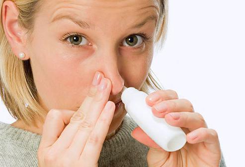
From an early age it is necessary to get used to the fact that the nose and the nasal cavity need regular hygiene. External respiratory passages must be cleaned of waste products, if necessary, you must also moisturize them. The same applies to periods of rhinitis: the blowing out of mucus must be done efficiently and carefully so that its particles do not fall into the passages connecting the nose to the ear.
As a rule, doctors talk about a large role in the prevention of sinusitis of such a simple measure as sanitation or washing the nasal cavity. This is not the most pleasant procedure, but it helps to get rid of pathogenic bacteria that have settled on the mucous membrane.


Popular
- Breast cancer is curable at any stage.
- The remedy for the cold Sinupret
- Azitrox - official instructions for use
- Chicken-bjaka: allowed antibiotics were found in Russian chicken
- Oral Cancer: Symptoms and Treatment
- Dark and thick blood during menstruation.
- Modern analogues of doxycycline tablets
- Is it possible to die from pneumonia
- What earwax will tell all about your health
- Tussin: instructions for use