Sinusitis - symptoms, diagnosis and treatment at home without a puncture. Use of antibiotics. Features of the structure of the nasal sinuses. Possible diseases
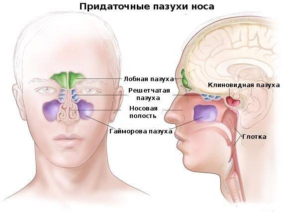
The mucous membrane of the nasal cavity and the sinuses are cleaned by the cilia located in them, and due to them the processes of moistening the cavity and its warming occur. In addition, they play the role of a voice resonator, which gives speech a peculiar shade, brightness and timbre. The near-nasal sinuses, or, as they are also called, adnexal, are special bone cavities in the skull, which are covered with mucous membranes and connected to the nose. A total of eight paranasal sinuses: maxillary or two maxillary, ethmoid labyrinths (front and rear), as well as two frontal and two wedge-shaped sinuses. It must be understood that the sinuses are filled with air, which subsequently goes out through the fistula. This is how the air exchange process or respiration process takes place in the body.
Nasal sinuses and their types
So, the following paranasal sinuses stand out:
- frontal;
- wedge-shaped;
- lattice labyrinth cells;
- maxillary.
 Air front with the nasal cavity is made through the middle nasal duct, and the rear is characterized by the upper nasal passage, which is important for the diagnosis of the disease of the posterior sinuses, especially the wedge-shaped ones, which are much less common than the front ones. Inside the sinuses have ciliated epithelium, with cells in the form of glasses and special mucus. Due to the active movement of the cilia, the mucus directly moves to the sinuses, with an approximate speed of 1 cm in one minute.
The structure of the nose and nasal cavity includes four types of sinuses, nasal conchas (middle and lower), nasal passage and nasal concha, pharyngeal mouth, soft palate, nasal vestibule and, of course, the tip of the nose. The nasal cavity itself is divided by a special septum, often into two halves, but with a curvature, it can also be divided into one.
The development of pathogens and diseases
The development of okolonosovy cavities begins in children, in fact, from birth, and they pumped their full formation to about 12 years. The most important for a person in medical terms are the maxillary sinuses, which are located on the sides of the nose, they are also called maxillary.
 In these sinuses, various inflammatory and infectious diseases can begin to develop; it is important to know and not to miss their development at an early stage. For example, inflammation of the sinuses can begin (with or without pus), which is called sinusitis or sinusitis.
In the paranasal sinuses may develop various pathological changes, for example, allergic reactions, polyps, cysts or congenital malformations. Of all the ENT organs, it is the diseases of the nasal sinuses that are considered the most dangerous and the most common. Among the inflammatory sinusitis of the nasal sinuses are the following forms:
- sinusitis, i.e. inflammation of the nasal sinuses, maxillary;
- ethmoiditis, i.e. diseases associated with labyrinths of the sinuses;
- frontal inflammation in the frontal cavities;
- sphenoiditis, sphenoid sinus disease.
The development of such diseases, due to the location of the paranasal groove, is very dangerous by the development of internal purulent accumulations, sometimes even several acute diseases are found at once.
Functions performed by the sinuses
The most common and important functions that the nasal sinuses perform are:

- decrease in the mass of the anterior parts of the skull, especially facial bone, according to their high volume;
- voice activation, tone brightness and coloring;
- protection of the roots of the teeth or eyeballs from exposure high temperatures, fluctuations from the breathing process;
- humidification of the air during inhalation and its warming;
- is an additional sense organ.
Development in the nasal cavity and sinuses acute diseases often begins against the background of the common cold, flu, in the presence of may be violations of air in the sinuses and a fairly rapid increase in the number of pathogens.
The reasons for which may cause sinusitis, are diseases of the teeth and gums, i.e. problems associated with the odontogenic area in, the result of such problems often becomes caries, periodontal disease, which border the lower wall of the maxillary sinuses, and can often have an effect on the sinuses. Acute odontogenic sinusitis quite often acquire a chronic form, due to the fact that they are very late to diagnose and treat.
With the manifestation of pathogenic organisms with effects on the nasal sinuses, the immunity is significantly reduced, the general background worsens, and the late treatment of such diseases can lead to the development of such a dangerous disease as diabetes, blood diseases and the gastrointestinal tract.
Symptoms that determine the appearance of pathologies of the paranasal sinuses are:

- blood or even purulent discharge from the nose;
- increase in human temperature, malaise, headache;
- nasal voice, timbre change;
- breathing problems;
- pain in the nasal cavity.
Diagnostic procedures of the nasal sinuses and treatment of the identified diseases should be carried out without fail by a specialized doctor who will professionally assess the integrity of the skull tissue, conduct an examination of the nasal passages using a rhinoscope. A nasal and oral swab will be required in order to clarify the characteristics of the contents and the identified disease.
In order to check the pathology of the nasal cavity, for a thorough check of the nasal secretions using the method of x-ray of the nasal cavity, CT and MRI.
Treatment and prevention of the development of pathology of the nasal sinuses are characterized depending on the form and type of the pathogen. For the treatment of acute forms may require surgery and classical methods of therapy. At the final stage of treatment, physiotherapy treatment is quite effective. It may even be worthwhile to use special, additional methods of treatment: sensing and washing.
Cleaning the sinuses and washing at home
In the case of a cold, you should take a closer look: if the painful condition and poor general body background lasts more than three days, it means that cold colds do not come out of the nasal cavity and appropriate measures should be taken to cleanse the sinuses and flush them.
The human body copes well with such microorganisms if they enter the body through swallowing or inspiration, but if they touch the sinuses, this can lead to serious infection and even death. Many experts recommend the use of cleansing, specially created for this liquid and drugs, use only sterile, distilled water. If, nevertheless, boiled water is used for washing, it must first be boiled for five minutes and be sure to cool to room temperature. Boiled water can be used to cleanse the sinuses during the day.
More effective methods can be used for washing and cleansing the paranasal sinuses, if the disease becomes more acute, for example, to purchase a special device in a pharmacy.
The most relevant among these devices are specialized kettles. These appliances, kettles are made of glass or ceramics and are often sold in pharmacies or health stores.
Rinse the nose with a solution of salt, stirred in warm distilled water. It is necessary to take about 5 tablespoons of salt (necessarily neiodarized so that there is no irritation) and, without boiling, heat and mix. Then you can rinse.
It is the washing of the nasal sinuses that will contribute to an effective and reliable cleansing of the cavity from mucus without the use of special chemicals and devices, but most importantly, it will allow you to breathe freely. But it is necessary to be extremely careful and attentive, because the sinuses of the nasal area are very sensitive and tender, therefore it is not recommended to use running water for cleansing, as it contains a large number of bacteria, microbes and various impurities.
Unlike popular belief, the nose is not only a “device” for breathing air. Inside the skull are the sinuses, which is a system connected by passages. They have the appearance of intracranial cavities and contain air. To find out what functions the nose and paranasal sinuses nose, called upper respiratory tract (upper DP), you need to consider what they are and what kind of structure they have.
Upper respiratory structure
The clinical anatomy of the nose and paranasal sinuses has a very complex structure. Directly, the nose serves as the beginning of the human respiratory system and continues through the cavity that connects the upper respiratory tract to the paranasal (paranasal) sinuses.
At the base of the external nose is a skeleton of bone and cartilage tissue covered with skin. All elements of the body are composed of cartilage and bone and skin. In turn, the base and back of the nose are composed of 3 types of bone elements, arranged in pairs. The cartilage system is also represented by three, arranged in pairs, species.
The space, called the nasal cavity, is located behind oral cavity and is divided into 2 halves by cartilaginous septum. It has 2 pairs of holes: front and rear. The anterior ones are called nostrils, and the posterior ones are called choans descending to the nasopharynx.
The nasal cavity is surrounded by the so-called paranasal sinuses. In medicine, they are divided into 4 pairs and can be: the frontal, sphenoid, maxillary and sinuses of the ethmoid bone. 
In addition, these cavities are located in the near and far parts of the skull. The course of pathologies in them is somewhat different. This is due to the fact that the anterior sinuses are connected to the nasal cavity by the middle one, and the back ones - by the upper nasal passage. At the same time, the incidence rate of the posterior sinuses is much lower than that in front.
A detailed diagram allows you to see in which area of the skull the sinuses are located.
Anatomy of the upper DP
The anatomical features of the paranasal sinuses are as follows:
The paranasal sinuses are supplied with blood from the ophthalmic and maxillary arteries. Their venous system is a type of extensive network branching in the area of natural fistulae. The outflow of blood through the nasal veins.
Features of the location of the sinuses in the child's body
The location of the paranasal sinuses in children differs significantly from the anatomical structure in adults. So, at birth, the baby has only 2 sinuses - the ethmoid and maxillary. In addition, both of them are represented by small diverticula of the nasal mucosa in the thickness of the bones. The maxillary sinus has a length of 10 mm, height and width - no more than 3 mm. Only by the 6th year of life the sinuses acquire normal forms, and at the age of 12 they descend to the location inherent in adults.
The rudiments of the ethmoid labyrinth of a newborn are located directly above the rudiments of the teeth. As the child grows, the teeth move to their natural place, and the sinus rises up and takes on the dimensions inherent in it. The main development of the ethmoid sinus begins at the age of 3 to 5 years. It is then that the number of cells and their size increases significantly.
The main sinus (wedge-shaped), like the frontal sinus, begins to form only in the fourth year of the baby’s life. By the age of 6, their sizes can be no more than 8x12 mm. Frequently, instead of two frontal sinuses only one develops or both are absent.
Types of inflammation in the sinuses
The structure of the nose and the paranasal cavities of the upper respiratory tract is such that they perform several important functions for the body. First of all, due to them the heat insulation of the brain and eyes is ensured, the mechanical strength of the bones of the skull increases. Sinuses also take part in voice formation and sound formation. But the main function of the organs is to clean, moisten and warm the air that enters the body from the external nose.
Since the paranasal sinuses are in direct contact with the air from the environment, they are prone to pathologies and inflammation caused by various causes. The most frequent pathogens of inflammation are:
- viral infections that enter the cavity through the nose, blood, etc .;
- bacteria and microorganisms: staphylococcus, fungi, etc.
In a healthy state, cilia of the epithelium, located in the nasal cavity and sinuses, remove mucus and dust particles and microorganisms to the outside. If this process is disturbed, inflammation may occur. Predisposing factors to the development of pathology are defects in the nasal septum and nasal shells, abnormal development of the upper PD, etc.
In addition, the causes of inflammation can be: 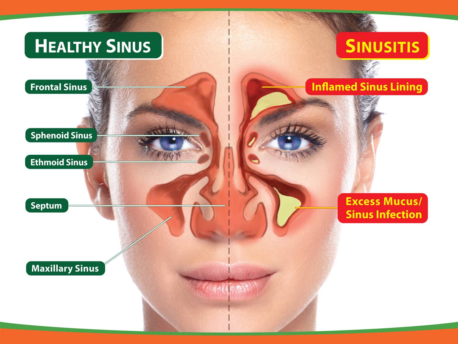
- injuries of the nose and paranasal sinuses;
- fever;
- inhalation of tobacco smoke and other harmful substances;
- hormonal disorders;
- low air humidity, etc.
Symptoms and signs of inflammation of the paranasal sinuses are quite characteristic.
An impulse to the development of the process can be a runny nose that has passed into rhinitis. The patient begins to complain of headaches, aggravated by bowing of the head or pressure drop, constant nasal congestion, temperature rise to 38 ° C and cough. Abundant and thick discharge greenish nose, bad breath, nasal voice.
The course of the disease may be acute or chronic. Acute inflammation, not burdened by complications, usually passes on its own within 14 days. In case of chronic course the process can be delayed for a long time, while the likelihood of recurrence is high.
Paranasal sinuses are quite fragile organs that require careful attention, especially at risk of developing complications. Even with modern medicine and equipment, doctors are not able to cope with some dangerous diseases. Despite this, timely diagnosis and therapeutic measures able to increase the chance of recovery.
In the front of the skull are cavities - voids, which are called paranasal sinuses. They perform the function of resonators, thanks to them the weight of the bones of the head is reduced. Each sinus with the nasal cavity is communicated through the fistula - a narrow connecting passage. There are several types of paranasal, or paranasal sinuses, differing from each other in location, size, structure.
Common to all paranasal sinuses
The anatomy of the nose and paranasal sinuses is especially actively formed during the first 5 years of life. Together with the nasal cavity, the paranasal sinuses constitute a single functional system.
All paranasal sinuses have walls that are dotted with numerous holes. Connective weaves, nerves go through these holes blood vessels. However, through the same holes in the cavity can penetrate:
- pus,
- toxins
- pathogenic flora,
- cancer cells with the spread in the orbits, pterygoid fossa, etc.
Due to the fact that the structure and physiology of the nose and the paranasal sinuses allows for the possibility of pathogen traffic, the development of secondary diseases and the occurrence of complications after, at first glance, non-dangerous infection of the individual sinuses are often observed.
Functions
One of the main tasks of the sinuses is to ensure the safety of the brain, orbits, facial nerves, arteries and veins. Anatomy of the paranasal sinuses normally suggests the possibility of unhindered withdrawal of constantly produced mucus, the physiological function of which is the neutralization of pathogens. The mucus is discharged along the fistulae, which must be opened for this, and is advanced to the exit thanks to the ciliated epithelium covered with multiple cilia.
With the onset of a cold, the production of mucus increases.
However, in case of significant edema of the mucous membrane and blockage of the fistula, exudate accumulates in the cavities. The reason for this may be:

In addition to the protective function are distinguished:
- resonant, due to which an individual voice timbre is formed,
- respiratory (in the process of nasal breathing, air circulates freely through the nasal passages, moistened and warmed),
- olfactory (the task is performed thanks to the recognition of smells of epithelial tissue).
Anatomical anomalies
The accessory sinuses of the nose differ in their diversity and their number and shape may vary in different people. For example, according to statistics, 5% of people are completely absent. In addition, topographic relationships, thickening or thinning of the walls of bone tissue, on the surface of which birth defects can also be, can be violated. Such anomalies occur in the late phase of prenatal (intrauterine) development.
Common anatomical anomalies include frontal and asymmetry. And to the rare - the complete absence of the maxillary cavity and the separation of the maxillary sinuses in half by bony septum.

This separation can occur both vertically (on the front and back), and horizontally (on the top and bottom).
More common cracking of the upper wall maxillary sinuswhich communicates with the infraorbital canal or the cavity of the orbit. The concavity of the facial wall in combination with the extension of the nasal wall into the lumen of the sinus threatens the penetration of the needle under the cheek when trying to puncture.
Anatomy and physiology depend on the genetic factor, which can be the cause of deformation of the facial and brain skeletons, as well as on the metabolism.
For all the sinuses in the paranasal region, the presence of slit paths of communication with the surrounding formations (dehiscence) is considered abnormal. For example, due to the occurrence of de-assessments:
- the ethmoid labyrinth is sometimes associated with the frontal and sphenoid sinuses, eye socket, cranial pits;
- the cracks in the side wall of the main sinus contribute to the contact of her mucosa with the dura (brain) of the middle cranial fossa, with the wing-palatal fossa, the superior orbital fissure and the optic nerve, the cavernous sinus and the internal carotid artery;
- thinning of the wall of the sphenoid sinus can lead to contact with the outlet and block nerves, with the branches of the oculomotor and trigeminal nerves.
Maxillary (maxillary) sinuses
Paired caves, which are located in the thickness of the bone. In an adult, the volume of each can reach 30 cm 3 (max), but the average volume is about 10 cm 3. In the form of a volume resembles a triangular pyramid. There are three of its walls:
- The upper (orbital) is the thinnest of the three, which is especially noticeable in its posterior part. Often in these places there are cracks, and sometimes completely absent bone tissue. Inside the wall from the infraorbital orifice passes the canal of the infraorbital nerve. If the canal is absent, the nerve and associated blood vessels are adjacent to the mucosa. However, in the event of inflammatory processes with such an arrangement, the probability of intraorbital and intracranial complications increases.
- The lower (cave bottom) is located near the back of the alveolar process (that is, near the upper jaw), so it sometimes happens that the sinus is separated from the four back upper teeth with only soft tissues. This proximity increases the risk due to odontogenic damage.
- The inner wall (also known as the lateral wall of the nasal cavity) normally corresponds to the middle and most of the lower nasal passages. In the back area of the lunate clippings under middle part nasal concha maxillary sinus through this wall opens with a hole in the nasal cavity. Everywhere, except for the lower parts, this wall is thin enough so that through it you can make therapeutic puncture.
Paired maxillary sinuses often differ in volume, with both shells (right and left) have bays (small additional depressions): alveolar, palatal, zygomatic, frontal.

Frontal (frontal) sinuses
They are paired cavities that are located in the thickness frontal bone, namely, between the plates of the scales and the orbital part. The right and left shells, as a rule, are separated by a thin partition. However, due to the nature of the formation, options are possible when:
- the partition is shifted to the left or right, which sometimes causes a significant difference in the size of the sinks,
- the septum may have openings that communicate between the frontal sinuses,
- cavities may be missing from one or both sides,
- the sinus may extend to the frontal scales, as well as to the base of the skull, along with the perforated plate of the ethmoid bone.
The frontal sinus communicates with the shell of the nasal cavity through the fronto-nasal canal. Its outlet is in the front of the mid-nasal passage.
The frontal shells become a continuation of the front cells of the ethmoid labyrinth, therefore in case of inflammation of one formation, the infection often spreads to another.
- Anterior wall - the place through which the piercing or opening of the sinus. Through the supraorbital cutting, the orbital nerve emerges.
- The lower wall is the thinnest of all that causes a simpler way of penetration of the infection into the orbit from the frontal shell.
- The brain wall, through which the infection can penetrate the anterior cranial fossa, it separates the shells from the frontal lobes.
Lattice labyrinth

The set of thin-walled cells consisting of bone tissue. Their average number is about 7-8 pieces, but the number can vary from 2 to 15. The cells are located in 3-4 rows, with conditional division into the front, rear and middle. They are located in the unpaired symmetrical ethmoid bone - in the frontal bone tenderloin. The posterior cells are in contact with the channel through which the optic nerve passes (sometimes it goes straight through them). Often the ethmoidal labyrinth reaches the most remote cavities of the facial skeleton, bordering on vital organs.
The labyrinth mucosa is innervated from the nasolabial nerve - the branch of the orbital nerve. In this regard, many diseases that occur with the defeat of the ethmoid labyrinth, are accompanied by pain sensations. Due to the fact that the olfactory filaments pass through tight canals of the skeletal bony plate, disturbances of smell are not uncommon in the development of edema due to squeezing.
Sphenoid (main) sinus
 Because of its location in the sphenoid bone (behind the lattice labyrinth above the arch of the nasopharynx and the joans), the main sinus has the second name, the sphenoid. In an adult, this sinus is divided into right and left non-communicating parts, which in most cases do not match in size and have independent exits to the nasal passage. Just describe the five walls of the cavity:
- Front. It consists of two parts: the nasal and lattice, which correlates with the back cells of the lattice labyrinth. The thinnest front wall flows smoothly into the lower one with the circulation in nasal cavity. In it there are small rounded holes through which the main sinus communicates with the nasopharynx. They are located at the level of the end of the upper shell of the nose.
- Back. Frontally located wall, less than a millimeter thick (with large volumes of the sinus), which causes the risk of damage to it during operations.
- Top. Corresponds to the bottom of the Turkish saddle, in which the optic nerve cross (enveloped in arachnoid membrane) and the pituitary gland are located. In the case of inflammation of the sphenoid sinus, it often goes to adjacent structures, sometimes affecting the olfactory tract or even the anteromedial surface of the frontal lobes of the brain.
- Lower Thick (about 12 mm) wall corresponding to the arch of the nasopharynx.
- Lateral. These walls border directly on the neurovascular bundles, which are located on the sides of the Turkish saddle. They can either absorb the channel of the optic nerve or come into contact with it. Through the wall on the border with the cavernous sinus and optic nerve infection can get into these structures.
Along with the listed sinuses, mention should be made of the pterygopal fossa, which is behind the tubercle. lower jaw. Its clinical significance is great because if inflammatory process nerves located in the fossa are involved, neuralgic syndromes of the facial part occur.
Sinus inflammation: types and symptoms
 Depending on the sinus in which the inflammatory process occurs, there are:
- sphenoiditis - inflammation affects the sphenoid sinus,
- sinusitis - affects the maxillary cavity,
- frontal disease - frontal zones are involved,
- ethmoiditis - the process takes place in the cells of the ethmoid labyrinth.
Inflammation of the mucous membranes can affect one or more sinuses at once. This inflammatory process occurs in different forms:
- acute form with pronounced symptoms
- recurrent - with less pronounced repetition of signs of acute inflammation,
- chronic.
The chronic form of the inflammatory process, which more often concerns the maxillary and slightly less often the frontal sinuses, lasts about 2-3 months, even if therapeutic measures are applied. Signs of a chronic process include:
- Discharge from the nose purulent, mucous, watery or mixed consistencies.
- Difficult breathing due to blockage of the nasal passages.
- Sore throat and reflex cough due to swelling of the mucous membranes at the back of the throat.
- Headaches occurring mainly in the nose, forehead and eyes.
- Violation of the olfactory function.
- The proliferation of polyps from the paranasal sinuses in the nasal passages.
Unlike children, adults are more likely to experience a viral infection of the nasal mucosa, which extends to the sinuses. Less commonly, the cause is blood diseases and dental health. The odontogenic factor is essential in the defeat of the maxillary sinuses. TO viral infection Against the background of the work of the "busy" immune system, the bacterial factor can join and become active - most often in the form of staphylococci.
Normally, microorganisms and microparticles, when inhaled, pass through the nasal cavity with air, enter the sinus caves, where the ciliated epithelium captures them and neutralizes them to form mucus. This mechanism can be disturbed by the curvature of various bone formations with anatomical deformation of the shells, as well as adverse factors affecting the protective properties of the epithelium: dry air, tobacco smoke, chemical burns, atrophy and necrosis of tissues, depressed state of the immune system, etc. Swelling may also occur as a result of an allergic reaction.
 Among the most common common symptoms of sinus inflammation are called:
- runny nose with thick greenish discharge and pus
- headache, which is aggravated by pressure drops, when the head is tilted, pressure is applied to areas in the nasal sinuses, as well as a feeling of distention in these areas,
- nasal congestion
- increase to 38C body temperature,
- morning and night cough.
Because of congestion, a person begins to breathe through his mouth, says nasal voices. At the same time, there is often an unpleasant smell from the mouth.
When sinusitis headaches associated with abnormal increase in intracranial pressure - one of the main signs. Pain in the forehead and sinuses may be pulsating or squeezing in nature, which is characteristic, first of all, for the acute form. In addition to the above signs noted:
- decrease in sense of smell (or loss of it),
- tearing and fear of light
- sometimes swelling of the upper eyelid or cheeks.
In the chronic course of the disease, the discharge flows down the pharyngeal wall, provoking a night cough. In the mornings and evenings there is a characteristic pain extending to the orbit area. When pressing on inner corner eye pain extends to the whole face.
Treatment of inflammation
Treatment of inflammation is carried out by conservative or surgical methods, depending on the evidence. Conservative methods involve the removal of edema of the mucous membranes, the destruction of pathogens, the creation of conditions for the removal of mucus and the organization of patency of the mouth of the sinus.
In the treatment of the acute form without the need to remove cysts, polyps, eliminate the curvature of the septum is used:
- vasoconstrictor - to relieve swelling,
- antibiotics local action - with purulent inflammation,
- antiseptic solutions in combination with washing through the puncture of the most convenient and thin wall,
- oil preparations for moisturizing dry mucous membranes, eliminating crusts,
- salt solutions during washing to moisten and normalize drainage of exudate.
Method "Cuckoo" with antritis
 Rinsing is applied only in the absence of disturbances in the structure of the fistula under the condition of normal circulation of fluid through the nasal cavity. It is performed without anesthesia. The patient lies on his back. A catheter is inserted into one nostril for delivering the medicine, and into the other a tube with a vacuum pump for pumping out fluid. In the course of the procedure, the patient pronounces the imitative imitation “ku-ku” which gives the name to the method in order to prevent the medication from getting through the throat into the respiratory tract. When the medication is applied, a slight pressure is created to facilitate the leaching of the exudate. In the treatment of sinusitis is usually prescribed 5 sessions.
Sometimes washing is combined with laser exposure, which is used to relieve swelling.
Sinus catheter flushing
Without a puncture, it is possible to treat sinusitis using the drug "Yamik." For flushing the patient, catheters are inserted through which high and low pressure is created (for this purpose, an air balloon is connected). Through one catheter, the contents of the sinuses are pumped out, and through the other the medicinal solution is supplied. The procedure is performed under local anesthesia.
Cyst
A cyst is detected by radiography. Without it, patients almost do not notice the neoplasm until it reaches a significant size comparable to the volume of the sinus. In this case, symptoms characteristic of sinusitis begin to manifest: headaches, a feeling of fullness, difficulty in nasal breathing. There is a cyst in violation of the ducts of the mucous gland, because of which the mucus is collected in a spherical capsule. Eliminated only surgically after determining its exact location using CT and MRI:
- The classic method involves the incision of the wall under the upper lip, which is associated with prolonged scarring and frequent subsequent recurrences of sinusitis.
- The endoscopic method is performed using an endoscope with a camera through the fistula, which eliminates traumatic complications.
Fungal infection
 Fungal inflammation is not considered rare. One nasal sinus is affected by the fungus or several at once.
People at risk of HIV infection and diabetics, and the probability of infection increases in people:
- conducting local treatment steroids
- taking antibiotics regularly,
- using drug therapy, leading to depression of the immune system,
- who underwent radiotherapy and chemotherapy for cancer.
The inflammatory reaction is most often provoked by the fungi of the Candida, Mukor, Aspergillus and Rhizopus genera.
At the same time, the symptoms of a fungal infection are similar to bacterial infection. The pattern of the disease can range from slow development to rapid growth of fungal growths with severe manifestations. An accurate diagnosis is established first with the help of radiological images, and then it is updated by conducting histological and mycological analyzes. In the case of fungal infection most often combine antifungal drug treatment with surgery aimed at removing polyps from the sinuses.
Features of inflammation in children
90% of all cases of inflammation of the nasal sinus in children is bacterial. Due to the fact that at this age there are a large number of variants of manifestations, sometimes there are difficulties with diagnosis. When inflammation in newborns for diagnosis, focus on:
- cough,
- smell from the mouth,
- switching to mouth breathing
- blocked nasal passages.
A specific sign can be attributed to the swelling of the eyelids and / or displacement towards the eyeball, which is associated with the location of the ethmoid sinus near the eye sockets, which in infants are separated from the sinus by a wall that is not yet fully formed. These manifestations are observed against the background of common symptoms: loss of appetite, tearfulness, worsening of sleep. Older children may additionally complain about the sensations of pain and pain in the area of the eyes. They also have nasal congestion, alternating with purulent-mucous secretions.
Before you start talking about sinusitis, as one of the complications of influenza or an independent disease, you must have an idea about the anatomy of human sinuses. This will help us photos and layout of the paranasal sinuses, below.
A person has 4 sinuses:
1) the maxillary or maxillary sinus (steam room, located on both sides of the nose);
2) paired frontal sinus;
3) the ethmoid labyrinth is also a sinus and also a steam room, and it is named this way because it is formed by the cells of the ethmoid bone and resembles a bone maze;
4) sphenoid sinus.
All the sinuses are important for humans, as they promote sound conduction, reduce the weight of the skull bones (the head would be too heavy if the bones of the skull were 100% filled with dense bone tissue), provide a shock-proof buffer for injuries. So to say that there would be no sinuses and there would not be the same sinusitis is fundamentally wrong, if the structure is present, it means it carries some useful function for a person.

The source of inflammation in the human body may be an infection that was able to undergo a protective formation and infectious agents begin reproduction in human tissues. Inflammation of one or more paranasal sinuses is called sinusitis. Depending on which of the sinuses has been attacked and the names of inflammation are distinguished:
1) antritis - with inflammation of the maxillary (maxillary) sinus;
2) frontal sinusitis - inflammation of the frontal sinus;
3) ethmoiditis - inflammation of the ethmoid cells;
4) sphenoiditis - inflammation of the sphenoid sinus.
Now that we’ve understood what sinuses and sinusitis are, let's talk specifically about the most frequent statistically sinus inflammation, sinusitis.
Etiology.
Sinusitis most often occurs on the background of infection in the maxillary sinus. Allergic sinusitis is very rare. An infection can penetrate into the sinus, either directly from the nose, or with blood flow can be brought into the sinus.
The infection can penetrate through the hole in the side wall of the nose, from which the mucous secretion that produces the epithelium of the sinus (it lines it from the inside) constantly enters the nasal cavity from the sinus with the help of flickering cilia. The role of this process is to keep the secret from fixing the infection on the epithelium. The mucus captures bacteria and then with the help of cilia the contaminated secret is carried into the nasal cavity through the opening. And when inflammation occurs, swelling of the nasal mucosa, the hole is blocked, mucus can not go into the nasal cavity and the sinus is not emptied, microbes begin to multiply inside the sinus, pus is formed, which leads to the development of the disease and symptoms of sinusitis. Pus can fill the entire bosom and even burst into the surrounding tissues, which is very dangerous symptom, as in this place is the concentration of vital human organs.
In addition to the structure of the nasal sinuses, the presence of anatomical defects or nasal polyps that prevent the secretions from escaping from the sinuses, one of the predisposing factors for the development of sinusitis may be a decrease in general immunity, which is observed during influenza infection. You can also note self-medication for catarrhal diseasessuch as inadequate use vasoconstrictor drops for the nose, which may even lead to a chronic process.
Children may have sinusitis even more often than adults because of the imperfection of the protective structures of the body, anatomical defects of the nasal wall and the presence of polyps.
Symptoms
1) pain. Initially, in the area of the affected sinus, then the pain tends to spread, so that the whole head starts to hurt. The pain most often intensifies in the evening. If sinusitis one-sided, it hurts more often that half of the head, where the inflamed sinus is located.
2) Nasal congestion. May be marked as unilateral congestion nasal and nasal breathing completely overlap. The voice of the diseased may acquire a nasal hue.
3) Runny nose. They can be either mucus from the nose (clear or yellowish), or purulent snot (yellow, green from the nose). Purulent crusts may form inside the nasal passages. Either runny nose may be absent altogether, this also happens.
4) Disturbance of smell and tearing - infrequent satellites of sinusitis, but also can help in the correct diagnosis, if they are present in the patient.
5) Temperature. It can reach up to 38 degrees, especially when sharp sinusor temperature may be absent.
6) General malaise. Weakness, sweating, fatigue due to general intoxication of the body can also occur with sinusitis.
Diagnostics.
It is possible to reliably determine whether a person has sinusitis or these are symptoms of other diseases using an x-ray. The study is called - X-ray examination of the paranasal sinuses. You can also carry out computed tomography of the sinuses, but this is not a way for mere mortals, given the cost of the method. X-rays, in most cases, are sufficient for correct diagnosis.
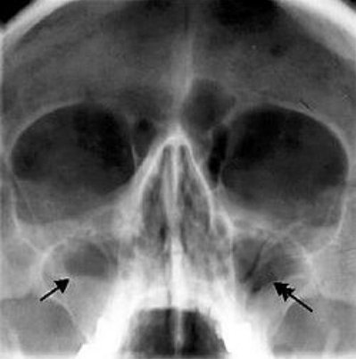
sinusitis with x-ray examination
Blood may show leukocytosis (an increase in the number of leukocytes) with a neutrophilic left shift (the medical term is to make it easier to understand this increase in the number of neutrophils, which are a form of leukocytes). All this is determined in general analysis blood.
Treatment.
The main purpose of the treatment of sinusitis is to eliminate the edema of the mucous holes between the maxillary sinus and the nasal cavity, since only in this case it is possible to remove pus from the cavity and improve the condition with subsequent recovery. The trouble is that inside the pus and bacteria themselves provoke swelling of the mucous membrane, thereby supporting the inflammatory process. Treatment should be carried out under the supervision of an ENT doctor.
All therapy includes conservative and surgical treatments.
Of conservative You can note the use of a variety of drugs and physiotherapy. This therapy can be performed at home and on an outpatient admission (in the clinic).
The first is the use of various drops, sprays and inhalers to eliminate swelling of the mucous. For this purpose, a whole layer of various vasoconstrictor drugs (Naphthyzinum, Galazolin, Nazol, Otrivin spray, which I like more than others on the effects and lack of side effects). It is only necessary to remember that it is necessary to bury the medicine for sinus correctly and do it on the side, so that the drug falls on the side wall of the nose, where the swollen opening of the entrance to the maxillary sinus is located.
After the introduction vasoconstrictor agents It is possible to use other drugs in drops: antibacterial (use antibiotics of the cephalosporins group), antihistamines.
Of the methods traditional medicine well helps washing the nasal cavity with a solution of salt. To do this, prepare saline solution: we take a teaspoon of salt to a glass of warm water, dissolve and retract this solution into each of the nostrils (only not hard, so that it does not fall deep) and blow our nose without effort, so that only the water and the pus that she took out. Repeat the procedure 5-7 times a day. The method is very similar to.
In addition, you can also use the heating of the sinuses with salt. I am telling how this can be done: you warm up salt in a dry frying pan, wrap the heated salt in a bag made of several layers of dense fabric and apply so that the heating of the maxillary sinuses takes place. Sometimes washing with salt and warming allow to achieve a phenomenal result in the treatment of sinusitis.
Of the specific drugs for the treatment of sinusitis is worth mentioning Sinuforte. At first, this drug inspired mistrust due to the aggressive advertising policy of the manufacturer, but then the results of this drug’s research began to appear in the international base, plus there was personal experience of using the drug for conservative (without a puncture) treatment of sinusitis. So I can say that the drug is worth it. The drug is based on chemical substances saponins, obtained from the juice of cyclamen roots, they have a local irritating effect on the nasal mucosa and contribute to the liquefaction of the viscous mucus layer. I will talk about this drug and its evidence base separately, since there is something to say, but you can trust this drug and its effectiveness has been confirmed, including in the patient feedback.
Ultraviolet irradiation of the nasal cavity (ultraviolet irradiation of the nose) and UHF on the paranasal sinuses (heating of the sinuses) are used as physiotherapy.
Surgical treatments.
From surgical methods of treatment it is worth noting the puncture of the maxillary sinus or puncture. Through the hole made by a special needle from the cavity maxillary sinus pus is removed, medications used for conservative therapy are also administered there. It turns out the delivery of drugs directly to the focus of inflammation. Patients notice relief after the first puncture. The method of treatment is not the most pleasant, from the side it looks even more unpleasant. During the treatment, several punctures have to be made, which makes a person painful due to the painful procedure. Currently, catheters are inserted into the puncture, which serve as a function of the emptying of the sinus and eliminating the need for a second puncture.
Of the myths, it is worth noting the steadiest, that if you made a puncture for sinus, then you will do it with every sinus inflammation, which is nonsense, since no direct correlation has been established.
But it is necessary to begin, as with any disease, from the simplest, with conservative therapy, since in most cases the correct combination of drugs helps to get rid of sinusitis without a puncture at home (on an outpatient basis).
Complications.
With the wrong treatment of sinusitis or lack of treatment, local and general complications can develop, such as:
- Chronization of the process with the development of chronic antritis;
- abscess or cellulitis of the orbit;
- intracranial abscess;
- Meningitis;
- sepsis.
Needless to say that delaying the treatment to the doctor or inadequate self-treatment can lead to disastrous consequences.
Prevention.
For the prevention of sinusitis using standard methods of prevention, as with any other infection. Take the same, the same methods for the overall strengthening of the body, plus add timely treatment infectious diseasesespecially related to diseases of the upper respiratory tract (flu, ARVI, bronchitis).
From specific prophylaxis in case of frequent sinusitis, it is worthwhile to approach the detection of anatomical defects of the nasal cavity and the nasal septum more carefully and correct them, including surgically.
Here it is such as sinusitis and as an independent disease, and as a complication after influenza infection. I would be glad if I could help and enlighten those in need.


Popular
- Breast cancer is curable at any stage.
- The remedy for the cold Sinupret
- Azitrox - official instructions for use
- Chicken-bjaka: allowed antibiotics were found in Russian chicken
- Oral Cancer: Symptoms and Treatment
- Dark and thick blood during menstruation.
- Modern analogues of doxycycline tablets
- Is it possible to die from pneumonia
- What earwax will tell all about your health
- Tussin: instructions for use